Navegação por Autores IPEN "ZEZELL, DENISE"
- Página inicial
- →
- Navegação por Autores IPEN
- Sobre
- Perfil Técnico
- Política de funcionamento
- Ajuda
- Apresentação
Navegação por Autores IPEN "ZEZELL, DENISE"
Itens para a visualização no momento 1-20 de 30
-
. Analysis of ceramic laminates removal with Er,Cr:YSGG laser by optical coherence tomography. Photobiomodulation, Photomedicine, and Laser Surgery, v. 37, n. 10, p. A22-A22, 2019. DOI: 10.1089/photob.2019.29013.abstracts Abstract: Porcelain laminated veneers have been widely used. For wear of hard tissue such as enamel and dentin, the diamond rotary instrument is the most traditional, but the laser has become recently used to remove aesthetic facets. Optical coherence tomography (OCT) used as an optical biopsy, is important for morphological analysis and attenuation coefficient is related to the property of the photons to be scattered by the samples. After approval by the Ethics Committee, the present study investigated the detachment of 30 ceramic E-max fragments cemented in human dental enamel of dimensions 3mm x 3mm x 0.7mm with 3 types of resin cements, RelxY Veneer, Relx U200 and Variolink Veneer. The samples (Enamel + Ceramic Fragment) were randomly distributed in the 3 groups and cemented according to the manufacturer. After that, they were prepared for irradiation with the Er,Cr: YSSG laser under predetermined conditions (3.5 and 3W, 20Hz, 60% water and 40% air flow). OCT analysis was done before and after irradiation. We observed that themorphological changes of the enamel surface showed an increased surface area due to the cement remaining in the enamel.We concluded that the Er, Cr: YSGG laser, when used in the irradiation protocol tested, seems to be a safe tool for the removal of laminates.. Analysis of ceramic laminates removal with Er,Cr:YSGG laser by optical coherence tomography. Photobiomodulation, Photomedicine, and Laser Surgery, v. 37, n. 10, p. A22-A22, 2019. DOI: 10.1089/photob.2019.29013.abstracts. Disponível em: http://repositorio.ipen.br/handle/123456789/32224. Acesso em: $DATA.Como referenciar este item
Esta referência é gerada automaticamente de acordo com as normas do estilo IPEN/SP (ABNT NBR 6023) e recomenda-se uma verificação final e ajustes caso necessário.
-
. Assessment of bound water of saliva samples by using FT-IR spectroscopy. In: LATIN AMERICA OPTICS AND PHOTONICS CONFERENCE, August 7-11, 2022, Recife, PE. Proceedings... Washington, DC, USA: Optica Publishing Group, 2022. DOI: 10.1364/LAOP.2022.M4B.1 Abstract: The objective of the present work is to show the relationship of the high-wavenumber spectral region concern OH vibrations, which show in a way how bound water can be altered in different sample groups.. Assessment of bound water of saliva samples by using FT-IR spectroscopy. In: LATIN AMERICA OPTICS AND PHOTONICS CONFERENCE, August 7-11, 2022, Recife, PE. Proceedings... Washington, DC, USA: Optica Publishing Group, 2022. DOI: 10.1364/LAOP.2022.M4B.1. Disponível em: http://repositorio.ipen.br/handle/123456789/33711. Acesso em: $DATA.Como referenciar este item
Esta referência é gerada automaticamente de acordo com as normas do estilo IPEN/SP (ABNT NBR 6023) e recomenda-se uma verificação final e ajustes caso necessário.
-
. Assessment of the optical attenuation coefficient of erored dentin. Lasers in Surgery and Medicine, v. 47, Suppl. 26, p. 32-33, 2015.
Palavras-Chave: coherent radiation; attenuation; dentin; neodymium lasers; fluorine; diagnostic uses; tomography; optical models
. Assessment of the optical attenuation coefficient of erored dentin. Lasers in Surgery and Medicine, v. 47, p. 32-33, 2015. Suppl. 26. Disponível em: http://repositorio.ipen.br/handle/123456789/23867. Acesso em: $DATA.Como referenciar este itemEsta referência é gerada automaticamente de acordo com as normas do estilo IPEN/SP (ABNT NBR 6023) e recomenda-se uma verificação final e ajustes caso necessário.
-
. Assessment to the optical attenuation coefficient of erored dentin. Lasers in Surgery and Medicine, v. 47, Sippl. 26, p. 32-33, 2015.
Palavras-Chave: attenuation; dentin; in vitro; evaluation; tomography; irradiation; neodymium lasers; fluorides; yttrium compounds
. Assessment to the optical attenuation coefficient of erored dentin. Lasers in Surgery and Medicine, v. 47, p. 32-33, 2015. Sippl. 26. Disponível em: http://repositorio.ipen.br/handle/123456789/26467. Acesso em: $DATA.Como referenciar este itemEsta referência é gerada automaticamente de acordo com as normas do estilo IPEN/SP (ABNT NBR 6023) e recomenda-se uma verificação final e ajustes caso necessário.
-
. Biochemical changes in normal skin caused by squamous cell carcinoma using FTIR spectroscopy. Lasers in Surgery and Medicine, v. 47, Suppl. 26, p. 2, 2015.
Palavras-Chave: carcinomas; skin; biological radiation effects; fourier transform spectrometers; infrared spectra; diagnostic uses
. Biochemical changes in normal skin caused by squamous cell carcinoma using FTIR spectroscopy. Lasers in Surgery and Medicine, v. 47, p. 2, 2015. Suppl. 26. Disponível em: http://repositorio.ipen.br/handle/123456789/23868. Acesso em: $DATA.Como referenciar este itemEsta referência é gerada automaticamente de acordo com as normas do estilo IPEN/SP (ABNT NBR 6023) e recomenda-se uma verificação final e ajustes caso necessário.
-
. Biochemical characterization of skin burn wound healing using ATR-FTIR. In: SBFOTON INTERNATIONAL OPTICS AND PHOTONICS CONFERENCE, October 08-10, 2018, Campinas, SP. Proceedings... Piscataway, NJ, USA: IEEE, 2018. DOI: 10.1109/SBFoton-IOPC.2018.8610943 Abstract: Efficient biochemical characterization of skin burn healing stages can improve clinical routine to adjust patients treatment. The golden standard for diagnosing skin burning stages is the histological biopsy. This practice is often expensive and technically challenging. There have been advances in the treatment, and diagnostic of the critical skin burned patients due to the increase of multidisciplinary collaboration. The contributions from different fields of biomedical engineering motivate to develop a better procedure for clinical applications. Considering the difficulty of monitoring wound healing the Fourier Transform Infrared coupled with an Attenuated Total Reflectance (ATR-FTIR) accessory is an analytical technique that can provide information regarding spectral biomarkers in biological materials. This study aimed to evaluate the classification feasibility provided by ATR-FTIR technique in the burned skin to follow the regenerative process in vivo. 40 skin burned samples from the Wistar rats dorsum at 3,7, 14, 21 days after burn were compared with the corresponded healthy group samples, by registering their infrared absorption spectra in FTIR Thermo Nicolet 6700 coupled to a diamond crystal ATR. The spectra were separated in the region 900 to 1800 cm-1 for further chemometric calculations. The second derivative of spectra was applied for discrimination, which results demonstrated differences from control and burns wounded groups, as well as among, burn wounded groups, using Amide I (1628 cm-1) and Amide II (1514 cm-1) bands. Amide I and Amide II bands are two significant bands of the infrared protein spectrum. The Amide I band is mainly associated with the C=O stretching vibration (70-80%) and is directly related to the backbone conformation. The Amide II band results from the N-H bending vibration (40-60%) and from the C-N stretching vibration (18-40%). This band is conformationally sensitive. These bands suggest proteins activity changing associate to inflammatory and maturation stages when it is compared with the healthy group. The statistical difference with amide I occur in proliferation and maturation stages. These findings indicate that ATR-FTIR is suitable to detect the burn wound healing stages and in the future can be an auxiliary instrument for clinical routine.. Biochemical characterization of skin burn wound healing using ATR-FTIR. In: SBFOTON INTERNATIONAL OPTICS AND PHOTONICS CONFERENCE, October 08-10, 2018, Campinas, SP. Proceedings... Piscataway, NJ, USA: IEEE, 2018. DOI: 10.1109/SBFoton-IOPC.2018.8610943. Disponível em: http://repositorio.ipen.br/handle/123456789/29822. Acesso em: $DATA.Como referenciar este item
Esta referência é gerada automaticamente de acordo com as normas do estilo IPEN/SP (ABNT NBR 6023) e recomenda-se uma verificação final e ajustes caso necessário.
-
. Calcium analysis from gamma sterilized human dentin and enamel. In: ENCONTRO DE OUTONO DA SOCIEDADE BRASILEIRA DE FÍSICA, 42., 26-31 de maio, 2019, Aracaju, SE. Resumo... São Paulo: Sociedade Brasileira de Física, 2019. Abstract: Gamma radiation changes the patients0 oral cavity undergoing radiotherapy. Alterations cause an unsaturated environment of calcium and phosphate into the oral cavity. After approval of the Ethics Committee, 20 hu- man teeth were sectioned to obtain 20 human enamel and 20 dentin samples, polished plane. Samples were randomized in the irradiated group and control group (untreated). Then, the treatment group was irradiated with 25:0 kGy at the 60Co multipurpose irradiator. After the gamma irradiation, Fourier Transformed Infrared Spectroscopy (FTIR), percentage of surface microhardness loss (%SMHL) and Scanning Electron Microscopy (SEM) were performed. At the end, acidic biopsies were performed to quantify the concentration of calcium present in the samples. FTIR showed that the molecular structure of HA of the enamel is similar to the non- irradiated, with no formation or loss of molecular compounds occurring. X-ray °uorescence at enamel samples was performed. Microscopic morphological analysis did not shown signi¯cant di®erences. Surface microhardness is an indirect indicator of the mineral content of the samples. The mean obtained was 258:2 (38:8) KHN within the hardness spectrum of the healthy natural enamel. The compounds present in the samples and the values of the ratios of Calcium and Phosphate oxides and relation between the elements Calcium and Phosphorus. The ratio of the most stable oxides shows a variation with linear correlation. In the enamel, the ratio (Ca/P) shows a change in the elemental content with linear correlation (R2 = 1). These ¯ndings lead us to a new hypothesis of behaviour of the HA crystal versus gamma irradiation. On the other hand for the irradiated dentin, the Knoop hardness number was within the range of the spectrum similar to that of natural dentin of human origin. X-ray °uorescence shows that irradiated dentin has great similarity with natural dentin from the point of view of chemical composition. SEM analyses showed that there was no thermal damage or interprismatic morpho- logical changes in the hydroxyapatite structure of human dental dentin outside the buccal environment when using doses of gamma irradiation up to 25 kGy.. Calcium analysis from gamma sterilized human dentin and enamel. In: ENCONTRO DE OUTONO DA SOCIEDADE BRASILEIRA DE FÍSICA, 42., 26-31 de maio, 2019, Aracaju, SE. Resumo... São Paulo: Sociedade Brasileira de Física, 2019. Disponível em: http://repositorio.ipen.br/handle/123456789/30214. Acesso em: $DATA.Como referenciar este item
Esta referência é gerada automaticamente de acordo com as normas do estilo IPEN/SP (ABNT NBR 6023) e recomenda-se uma verificação final e ajustes caso necessário.
-
. Can high power laser on swine mitral valve chordae tendineae improve mitral regurgitation? insights for a new surgical ery technique. In: ANNUAL CONFERENCE OF AMERICAN SOCIETY FOR LASER MEDICINE AND SURGERY, 32rd, April 3-7, 2013, Boston, MA, USA. Abstract... 2013. p. 64-65.
Palavras-Chave: heart; valves; rheumatic diseases; fever; surgery; corrections
. Can high power laser on swine mitral valve chordae tendineae improve mitral regurgitation? insights for a new surgical ery technique. In: ANNUAL CONFERENCE OF AMERICAN SOCIETY FOR LASER MEDICINE AND SURGERY, 32rd, April 3-7, 2013, Boston, MA, USA. Abstract... 2013. p. 64-65. Disponível em: http://repositorio.ipen.br/handle/123456789/20231. Acesso em: $DATA.Como referenciar este itemEsta referência é gerada automaticamente de acordo com as normas do estilo IPEN/SP (ABNT NBR 6023) e recomenda-se uma verificação final e ajustes caso necessário.
-
. Characterization of irradiated bone by FTIR. Lasers in Surgery and Medicine, v. Suppl.21, p. 78-79, 2009.
Palavras-Chave: bone tissues; chemical radiation effects; post-irradiation examination; fourier transform spectrometers; infrared spectra; surgery; laser radiation
. Characterization of irradiated bone by FTIR. Lasers in Surgery and Medicine, v. Suppl.21, p. 78-79, 2009. Disponível em: http://repositorio.ipen.br/handle/123456789/8875. Acesso em: $DATA.Como referenciar este itemEsta referência é gerada automaticamente de acordo com as normas do estilo IPEN/SP (ABNT NBR 6023) e recomenda-se uma verificação final e ajustes caso necessário.
-
. Chemometric methods applied to FTIR spectra to discriminate treated and non-treated cutaneous malignant lesions from healthy skin. In: LATIN AMERICA OPTICS AND PHOTONICS CONFERENCE, August 22-26, 2016, Medellín, Colombia. Proceedings... Washington, DC, USA: OSA, 2016. Abstract: Chemometric methods were used to differentiate FTIR spectra of treated and nontreated malignant lesions from healthy skin. We conclude that the method can be used to evaluate the biological changes promoted by photodynamic treatment.
Palavras-Chave: skin; formation damage; melanomas; chemotherapy; photodynamic therapy
. Chemometric methods applied to FTIR spectra to discriminate treated and non-treated cutaneous malignant lesions from healthy skin. In: LATIN AMERICA OPTICS AND PHOTONICS CONFERENCE, August 22-26, 2016, Medellín, Colombia. Proceedings... Washington, DC, USA: OSA, 2016. Disponível em: http://repositorio.ipen.br/handle/123456789/27032. Acesso em: $DATA.Como referenciar este itemEsta referência é gerada automaticamente de acordo com as normas do estilo IPEN/SP (ABNT NBR 6023) e recomenda-se uma verificação final e ajustes caso necessário.
-
. Comportamento da hidroxiapatita do esmalte e da dentina frente à radiação ionizante in vivo e in vitro. In: CONGRESSO UNIVERSITÁRIO BRASILEIRO DE ODONTOLOGIA, 43., 18-20 de setembro, 2019, São Paulo, SP. Resumo expandido... 2019.. Comportamento da hidroxiapatita do esmalte e da dentina frente à radiação ionizante in vivo e in vitro. In: CONGRESSO UNIVERSITÁRIO BRASILEIRO DE ODONTOLOGIA, 43., 18-20 de setembro, 2019, São Paulo, SP. Resumo expandido... 2019. Disponível em: http://200.136.52.105/handle/123456789/31783. Acesso em: $DATA.Como referenciar este item
Esta referência é gerada automaticamente de acordo com as normas do estilo IPEN/SP (ABNT NBR 6023) e recomenda-se uma verificação final e ajustes caso necessário.
-
. Descriptive analysis of in vitro cutting of swine mitral cusps: comparison of high-power laser and scalpel blade cutting techniques. Photomedicine and Laser Surgery, v. 35, n. 2, p. 87-91, 2017. DOI: 10.1089/pho.2015.3993 Abstract: Background and objectives: The most common injury to the heart valve with rheumatic involvement is mitral stenosis, which is the reason for a big number of cardiac operations in Brazil. Commissurotomy is the traditional technique that is still widely used for this condition, although late postoperative restenosis is concerning. This study's purpose was to compare the histological findings of porcine cusp mitral valves treated in vitro with commissurotomy with a scalpel blade to those treated with high-power laser (HPL) cutting, using appropriate staining techniques. Materials and methods: Five mitral valves from healthy swine were randomly divided into two groups: Cusp group (G1), cut with a scalpel blade (n = 5), and Cusp group (G2), cut with a laser (n = 5). G2 cusps were treated using a diode laser (lambda = 980 nm, power = 9.0 W, time = 12 sec, irradiance = 5625 W/cm(2), and energy = 108 J). Results: In G1, no histological change was observed in tissue. A hyaline basophilic aspect was focally observed in G2, along with a dark red color on the edges and areas of lower birefringence, when stained with hematoxylin-eosin, Masson's trichrome, and Sirius red. Further, the mean distances from the cutting edge in cusps submitted to laser application and stained with Masson's trichrome and Sirius red were 416.7 and 778.6 mu m, respectively, never overcoming 1mm in length. Conclusions: Thermal changes were unique in the group submitted to HPL and not observed in the cusp group cut with a scalpel blade. The mean distance of the cusps' collagen injury from the cutting edge was less than 1mm with laser treatment. Additional studies are needed to establish the histological evolution of the laser cutting and to answer whether laser cutting may avoid valvular restenosis better than blade cutting.
Palavras-Chave: cusped geometries; blood circulation; cardiovascular diseases; cardiovascular system; cutting; laser power transmission
. Descriptive analysis of in vitro cutting of swine mitral cusps: comparison of high-power laser and scalpel blade cutting techniques. Photomedicine and Laser Surgery, v. 35, n. 2, p. 87-91, 2017. DOI: 10.1089/pho.2015.3993. Disponível em: http://repositorio.ipen.br/handle/123456789/27894. Acesso em: $DATA.Como referenciar este itemEsta referência é gerada automaticamente de acordo com as normas do estilo IPEN/SP (ABNT NBR 6023) e recomenda-se uma verificação final e ajustes caso necessário.
-
. Discrimination of healthy skin and cutaneous malignant lesions using FTIR spectra and their second derivatives: a comparative study. In: CLINICAL AND TRANSLATIONAL BIOPHOTONICS, April 3-6, 2018, Hollywood, Florida, United States. Resumo expandido... Washington, DC, USA: OSA, 2018. Abstract: PC-LDA statistical method was used to differentiate cutaneous tumor tissue from healthy skin. Discrimination accuracy obtained by raw FTIR spectra was 95% and by second derivatives 92%, besides identifying secondary structure of proteins and collagen.. Discrimination of healthy skin and cutaneous malignant lesions using FTIR spectra and their second derivatives: a comparative study. In: CLINICAL AND TRANSLATIONAL BIOPHOTONICS, April 3-6, 2018, Hollywood, Florida, United States. Resumo expandido... Washington, DC, USA: OSA, 2018. Disponível em: http://repositorio.ipen.br/handle/123456789/29119. Acesso em: $DATA.Como referenciar este item
Esta referência é gerada automaticamente de acordo com as normas do estilo IPEN/SP (ABNT NBR 6023) e recomenda-se uma verificação final e ajustes caso necessário.
-
. Effects of lasers on chemical composition of anamel and dentin. In: ANNUAL CONFERENCE OF AMERICAN SOCIETY FOR LASER MEDICINE AND SURGERY, 29th, April 1-5, 2009, Washington. Abstract... 2009.
Palavras-Chave: dentin; enamels; laser radiation; morphological changes
. Effects of lasers on chemical composition of anamel and dentin. In: ANNUAL CONFERENCE OF AMERICAN SOCIETY FOR LASER MEDICINE AND SURGERY, 29th, April 1-5, 2009, Washington. Abstract... 2009. Disponível em: http://repositorio.ipen.br/handle/123456789/22838. Acesso em: $DATA.Como referenciar este itemEsta referência é gerada automaticamente de acordo com as normas do estilo IPEN/SP (ABNT NBR 6023) e recomenda-se uma verificação final e ajustes caso necessário.
-
. Effects of lasers on chemical composition on enamel and dentin. Lasers in Surgery and Medicine, v. Suppl.21, p. 53, 2009.
Palavras-Chave: enamels; dentin; laser radiation; radiation effects; morphological changes
. Effects of lasers on chemical composition on enamel and dentin. Lasers in Surgery and Medicine, v. Suppl.21, p. 53, 2009. Disponível em: http://repositorio.ipen.br/handle/123456789/8863. Acesso em: $DATA.Como referenciar este itemEsta referência é gerada automaticamente de acordo com as normas do estilo IPEN/SP (ABNT NBR 6023) e recomenda-se uma verificação final e ajustes caso necessário.
-
. Evaluation of calcified mitral valves after Er,Cr:YSGG irradiation using Optical Coherence Tomography. In: INTERNATIONAL CONFERENCE ON LASER APPLICATIONS IN LIFE SCIENCES, 16th, April 1-2, 2022, Nancy, France. Abstract... Nancy, France: PROGEPI, 2022. p. 152-152. Abstract: Mitral valve is responsible to control the left atrium-ventricle blood flux. Mitral stenosis is a disease that occurs in consequence of calcification and fibrosis on the cuspids of the valve. Diagnosis can be performed using echocardiography.Many treatments are possible, and one of them is commissurotomy (surgical approach).High intensity laser irradiation may be a new strategy for this surgical technique[1], and the optical coherence tomography (OCT) may contribute to the valve evaluation[2], asit provides higherspatialresolutionin exchange of lower penetrationthan ultrasonography. In this way, the aim of this study is to evaluate laser irradiation effectsincalcified mitral valvesusing OCTand digital processing.To that, it was conducted an ex-vivostudywith four human mitral valvessamples,obtained from valve replacement surgeries in the Heart Institute.The samples were splitin four groups: scalpel cut, laser cut, scalpel debridement and laser debridement.Cutting and debridement procedures were performed in calcified regions of the valves, usinga disposable scalpelbladeand anEr,Cr:YSGG laser(Waterlase; Biolase Inc., CA, USA), emitting at 2780 nm. The laser parameters were set at power = 1.6W, frequency = 20 Hz, energy density = 28.3J/cm2,pulse duration = 700 μs, 15% of water and 15% of air.The imaging was performed using a spectraldomain OCT system(Callisto110C1;ThorLabs Inc., NJ, USA).It was acquired10 B-scans per sample, 5 inprocedures regions and 5 in sound regions. The Optical Attenuation Coefficient (OAC) was calculated by comparing a beer-lambert like equation to exponential fittings of the A-scans[3].The distribution and normality of variances were tested using Shapiro-Wilk test,and statistical comparison was performed using one-way ANOVA and Tukey’s post hoc. All tests considered a level of significance of 5%.The FigureAshows a representative B-scan of a visibly calcified region, where a pattern of higher intensities can be observed.Thispattern is related tomorphological and optical changes, mainly a refractive index change, due to calcium presence in the valve tissue.This B-scan was acquired only to understand the calcified tissue aspect, as the procedures regions does notpresent visibly largecalcium stones.The Figure Bshowsthe statisticalanalysis, where the sound OAC values, as a mean of all sound regions, presented a significant statistical difference in comparison to scalpel groups, while no difference waspresentedin relation to laser groups. Higher OAC values are related to anaugmentation of the light backscatteringdue to calcium refractive index, leading to a change of lightpropagation in tissue-calcium interfaces.This finding indicates thatthe laser procedures promoted a better removal of calcified tissue than the scalpelmethods, which can be related to tissue-ablation interaction.Furthermore, the statistical difference between scalpel cut group and both laser groups suggests that the scalpel needs more wear interaction with the tissue, such as in the debridement procedure, being unable to significatively remove the calcification in a single cut.This study points the Er,Cr:YSGG and the OCT as potential techniques for the calcified tissue removal and evaluation,respectively, duringmitral valvessurgeries, although further studieswith higher sample numbermust be performed.
Palavras-Chave: cardiovascular system; valves; cardiovascular diseases; fibrosis; tomography; coherent radiation
. Evaluation of calcified mitral valves after Er,Cr:YSGG irradiation using Optical Coherence Tomography. In: INTERNATIONAL CONFERENCE ON LASER APPLICATIONS IN LIFE SCIENCES, 16th, April 1-2, 2022, Nancy, France. Abstract... Nancy, France: PROGEPI, 2022. p. 152-152. Disponível em: http://repositorio.ipen.br/handle/123456789/33940. Acesso em: $DATA.Como referenciar este itemEsta referência é gerada automaticamente de acordo com as normas do estilo IPEN/SP (ABNT NBR 6023) e recomenda-se uma verificação final e ajustes caso necessário.
-
. Evaluation of thermal stability of human dental tissue using fourier transform infrared spectroscopy. In: ENCONTRO NACIONAL DE FISICA DA MATERIA CONDENSADA, 32., 11-15 de maio, 2009, Aguas de Lindoia, SP. Resumo... 2009.
Palavras-Chave: laser radiation; teeth; animal tissues; temperature dependence; morphological changes; fourier transform spectrometers
. Evaluation of thermal stability of human dental tissue using fourier transform infrared spectroscopy. In: ENCONTRO NACIONAL DE FISICA DA MATERIA CONDENSADA, 32., 11-15 de maio, 2009, Aguas de Lindoia, SP. Resumo... 2009. Disponível em: http://repositorio.ipen.br/handle/123456789/19322. Acesso em: $DATA.Como referenciar este itemEsta referência é gerada automaticamente de acordo com as normas do estilo IPEN/SP (ABNT NBR 6023) e recomenda-se uma verificação final e ajustes caso necessário.
-
. FTIR hyperspectral imaging for label-free histopathology. In: INTERNATIONAL SYMPOSIUM AND INTERNATIONAL SCHOOL FOR YOUNG SCIENTISTS ON “PHYSICS, ENGINEERING AND TECHNOLOGIES FOR BIOMEDICINE”, 5th, November 21-25, 2020, Moscow, Russia. Abstract... Moscow, Russia: MEPhI, 2020. p. 65-65. Abstract: FTIR hyperspectral pathology imaging of thin tissue slice samples are used to monitor the collagen during the healing process when evalu-ating burned skin, treated or not with femtosecond laser, in the diagnose and molecular differentiation between thyroid and goiter, skin squamous cell carcinoma, as well as for breast cancer cell subtypes.. FTIR hyperspectral imaging for label-free histopathology. In: INTERNATIONAL SYMPOSIUM AND INTERNATIONAL SCHOOL FOR YOUNG SCIENTISTS ON “PHYSICS, ENGINEERING AND TECHNOLOGIES FOR BIOMEDICINE”, 5th, November 21-25, 2020, Moscow, Russia. Abstract... Moscow, Russia: MEPhI, 2020. p. 65-65. Disponível em: http://repositorio.ipen.br/handle/123456789/33358. Acesso em: $DATA.Como referenciar este item
Esta referência é gerada automaticamente de acordo com as normas do estilo IPEN/SP (ABNT NBR 6023) e recomenda-se uma verificação final e ajustes caso necessário.
-
. FTIR imaging on glass substrates evaluation of histological skin burn injuries specimens treated by femtosecond laser pulses. In: INTERNATIONAL CONFERENCE ON LASER APPLICATIONS IN LIFE SCIENCES, 16th, April 1-2, 2022, Nancy, France. Abstract... Nancy, France: PROGEPI, 2022. p. 205-205. Abstract: Burn injuries continue to be one of the leading causes of unintentional death and injury in low- and middle-income countries [1]. Burns are considered an important public health problem, because in addition to physical problems that can lead the patient to death, they cause psychological and social damage. An estimated 180,000 deaths every year are caused by burns [2]. The use of infrared (IR) spectroscopy for studying biological specimens is nowadays a wide and active area of research. The IR microspectroscopy has proved to be an ideal tool for investigating the biochemical composition of biological samples at the microscopic scale, as well as its fast, sensitive, and label-free nature [3]. IR image spectral histopathology has shown great promise as an important diagnostic tool, with the potential to complement current pathological methods, reducing subjectivity in biopsy samples analysis. However, the use of IR transmissive substrates which are both fragile and prohibitively very expensive, hinder the clinical translation. The goal of this study is to evaluate the potential of discriminating healing process, in burned skin specimens treated with ultrashort pulses laser 3 days after the burn. This study is considering a previous paper [4], in which it analyzed only micro-ATR-FTIR spectra of a frozen sample point. The specimens were obtained from third degree burn wound. The wounds treatment were performed three days after the burn, and the animals were sacrificed 3 and 14 days post-treatment. Using coverslipped H&E stained tissue on glass from previous histopathological analysis and applying the analytical techniques PCA and K-means on N−H, O−H, and C−H stretching regions occurring at 2500−3800 cm−1 (high wavenumber region), were possible to discriminate burned epidermal and dermal regions from irradiated in same regions on sample. In the figures is shown the average spectrum at (a) day 3 and (b) day 14. , in both there were increase of burned+laser treated bands. The great potential of this study was to analyse coverslipped H&E stained tissue on glass, without compromising the histopathologist practices and contribute for clinical translation.
Palavras-Chave: burns; injuries; animal tissues; infrared spectra; healing; lasers; pulses
. FTIR imaging on glass substrates evaluation of histological skin burn injuries specimens treated by femtosecond laser pulses. In: INTERNATIONAL CONFERENCE ON LASER APPLICATIONS IN LIFE SCIENCES, 16th, April 1-2, 2022, Nancy, France. Abstract... Nancy, France: PROGEPI, 2022. p. 205-205. Disponível em: http://repositorio.ipen.br/handle/123456789/33939. Acesso em: $DATA.Como referenciar este itemEsta referência é gerada automaticamente de acordo com as normas do estilo IPEN/SP (ABNT NBR 6023) e recomenda-se uma verificação final e ajustes caso necessário.
-
. FTIR microspectroscopy discriminating skin cancer using tissue sections on glass. In: CONFERENCE SPEC, 10th, June 10-15, 2018, Glasgow, UK. Resumo expandido... Manchester, UK: The International Society for Clinical Spectroscopy, 2018. p. 113-114.. FTIR microspectroscopy discriminating skin cancer using tissue sections on glass. In: CONFERENCE SPEC, 10th, June 10-15, 2018, Glasgow, UK. Resumo expandido... Manchester, UK: The International Society for Clinical Spectroscopy, 2018. p. 113-114. Disponível em: http://repositorio.ipen.br/handle/123456789/29823. Acesso em: $DATA.Como referenciar este item
Esta referência é gerada automaticamente de acordo com as normas do estilo IPEN/SP (ABNT NBR 6023) e recomenda-se uma verificação final e ajustes caso necessário.
Itens para a visualização no momento 1-20 de 30
Buscar no repositório
Navegar
Minha conta
Visualizar
A pesquisa no RD utiliza os recursos de busca da maioria das bases de dados. No entanto algumas dicas podem auxiliar para obter um resultado mais pertinente.
✔ É possível efetuar a busca de um autor ou um termo em todo o RD, por meio do Buscar no Repositório , isto é, o termo solicitado será localizado em qualquer campo do RD. No entanto esse tipo de pesquisa não é recomendada a não ser que se deseje um resultado amplo e generalizado.
✔ A pesquisa apresentará melhor resultado selecionando um dos filtros disponíveis em Navegar
✔ Os filtros disponíveis em Navegar tais como: Coleções, Ano de publicação, Títulos, Assuntos, Autores, Revista, Tipo de publicação são autoexplicativos. O filtro, Autores IPEN apresenta uma relação com os autores vinculados ao IPEN; o ID Autor IPEN diz respeito ao número único de identificação de cada autor constante no RD e sob o qual estão agrupados todos os seus trabalhos independente das variáveis do seu nome; Tipo de acesso diz respeito à acessibilidade do documento, isto é , sujeito as leis de direitos autorais, ID RT apresenta a relação dos relatórios técnicos, restritos para consulta das comunidades indicadas.

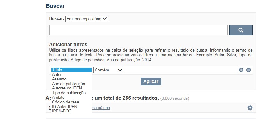
A opção Busca avançada utiliza os conectores da lógica boleana, é o melhor recurso para combinar chaves de busca e obter documentos relevantes à sua pesquisa, utilize os filtros apresentados na caixa de seleção para refinar o resultado de busca. Pode-se adicionar vários filtros a uma mesma busca.
Exemplo:
Buscar os artigos apresentados em um evento internacional de 2015, sobre loss of coolant, do autor Maprelian.
Autor: Maprelian
Título: loss of coolant
Tipo de publicação: Texto completo de evento
Ano de publicação: 2015

✔ Para indexação dos documentos é utilizado o Thesaurus do INIS, especializado na área nuclear e utilizado em todos os países membros da International Atomic Energy Agency – IAEA , por esse motivo, utilize os termos de busca de assunto em inglês; isto não exclui a busca livre por palavras, apenas o resultado pode não ser tão relevante ou pertinente.
✔ 95% do RD apresenta o texto completo do documento com livre acesso, para aqueles que apresentam o ![]() significa que e o documento está sujeito as leis de direitos autorais, solicita-se nesses casos contatar a Biblioteca do IPEN,
bibl@ipen.br
.
significa que e o documento está sujeito as leis de direitos autorais, solicita-se nesses casos contatar a Biblioteca do IPEN,
bibl@ipen.br
.
✔ Ao efetuar a busca por um autor o RD apresentará uma relação de todos os trabalhos depositados no RD. No lado direito da tela são apresentados os coautores com o número de trabalhos produzidos em conjunto bem como os assuntos abordados e os respectivos anos de publicação agrupados.
✔ O RD disponibiliza um quadro estatístico de produtividade, onde é possível visualizar o número dos trabalhos agrupados por tipo de coleção, a medida que estão sendo depositados no RD.
✔ Na página inicial nas referências são sinalizados todos os autores IPEN, ao clicar nesse símbolo ![]() será aberta uma nova página correspondente à aquele autor – trata-se da página do pesquisador.
será aberta uma nova página correspondente à aquele autor – trata-se da página do pesquisador.
✔ Na página do pesquisador, é possível verificar, as variações do nome, a relação de todos os trabalhos com texto completo bem como um quadro resumo numérico; há links para o Currículo Lattes e o Google Acadêmico ( quando esse for informado).
ATENÇÃO!
ESTE TEXTO "AJUDA" ESTÁ SUJEITO A ATUALIZAÇÕES CONSTANTES, A MEDIDA QUE NOVAS FUNCIONALIDADES E RECURSOS DE BUSCA FOREM SENDO DESENVOLVIDOS PELAS EQUIPES DA BIBLIOTECA E DA INFORMÁTICA.
O gerenciamento do Repositório está a cargo da Biblioteca do IPEN. Constam neste RI, até o presente momento 20.950 itens que tanto podem ser artigos de periódicos ou de eventos nacionais e internacionais, dissertações e teses, livros, capítulo de livros e relatórios técnicos. Para participar do RI-IPEN é necessário que pelo menos um dos autores tenha vínculo acadêmico ou funcional com o Instituto. Nesta primeira etapa de funcionamento do RI, a coleta das publicações é realizada periodicamente pela equipe da Biblioteca do IPEN, extraindo os dados das bases internacionais tais como a Web of Science, Scopus, INIS, SciElo além de verificar o Currículo Lattes. O RI-IPEN apresenta também um aspecto inovador no seu funcionamento. Por meio de metadados específicos ele está vinculado ao sistema de gerenciamento das atividades do Plano Diretor anual do IPEN (SIGEPI). Com o objetivo de fornecer dados numéricos para a elaboração dos indicadores da Produção Cientifica Institucional, disponibiliza uma tabela estatística registrando em tempo real a inserção de novos itens. Foi criado um metadado que contém um número único para cada integrante da comunidade científica do IPEN. Esse metadado se transformou em um filtro que ao ser acionado apresenta todos os trabalhos de um determinado autor independente das variáveis na forma de citação do seu nome.
A elaboração do projeto do RI do IPEN foi iniciado em novembro de 2013, colocado em operação interna em julho de 2014 e disponibilizado na Internet em junho de 2015. Utiliza o software livre Dspace, desenvolvido pelo Massachusetts Institute of Technology (MIT). Para descrição dos metadados adota o padrão Dublin Core. É compatível com o Protocolo de Arquivos Abertos (OAI) permitindo interoperabilidade com repositórios de âmbito nacional e internacional.
1. Portaria IPEN-CNEN/SP nº 387, que estabeleceu os princípios que nortearam a criação do RDI, clique aqui.
2. A experiência do Instituto de Pesquisas Energéticas e Nucleares (IPEN-CNEN/SP) na criação de um Repositório Digital Institucional – RDI, clique aqui.
O Repositório Digital do IPEN é um equipamento institucional de acesso aberto, criado com o objetivo de reunir, preservar, disponibilizar e conferir maior visibilidade à Produção Científica publicada pelo Instituto, desde sua criação em 1956.
Operando, inicialmente como uma base de dados referencial o Repositório foi disponibilizado na atual plataforma, em junho de 2015. No Repositório está disponível o acesso ao conteúdo digital de artigos de periódicos, eventos, nacionais e internacionais, livros, capítulos, dissertações, teses e relatórios técnicos.
A elaboração do projeto do RI do IPEN foi iniciado em novembro de 2013, colocado em operação interna em julho de 2014 e disponibilizado na Internet em junho de 2015. Utiliza o software livre Dspace, desenvolvido pelo Massachusetts Institute of Technology (MIT). Para descrição dos metadados adota o padrão Dublin Core. É compatível com o Protocolo de Arquivos Abertos (OAI) permitindo interoperabilidade com repositórios de âmbito nacional e internacional.
O gerenciamento do Repositório está a cargo da Biblioteca do IPEN. Constam neste RI, até o presente momento 20.950 itens que tanto podem ser artigos de periódicos ou de eventos nacionais e internacionais, dissertações e teses, livros, capítulo de livros e relatórios técnicos. Para participar do RI-IPEN é necessário que pelo menos um dos autores tenha vínculo acadêmico ou funcional com o Instituto. Nesta primeira etapa de funcionamento do RI, a coleta das publicações é realizada periodicamente pela equipe da Biblioteca do IPEN, extraindo os dados das bases internacionais tais como a Web of Science, Scopus, INIS, SciElo além de verificar o Currículo Lattes. O RI-IPEN apresenta também um aspecto inovador no seu funcionamento. Por meio de metadados específicos ele está vinculado ao sistema de gerenciamento das atividades do Plano Diretor anual do IPEN (SIGEPI). Com o objetivo de fornecer dados numéricos para a elaboração dos indicadores da Produção Cientifica Institucional, disponibiliza uma tabela estatística registrando em tempo real a inserção de novos itens. Foi criado um metadado que contém um número único para cada integrante da comunidade científica do IPEN. Esse metadado se transformou em um filtro que ao ser acionado apresenta todos os trabalhos de um determinado autor independente das variáveis na forma de citação do seu nome.
