Navegação IPEN por Autores IPEN "DEL-VALLE, MATHEUS"
- Página inicial
- →
- IPEN
- →
- Navegação IPEN por Autores IPEN
- Sobre
- Perfil Técnico
- Política de funcionamento
- Ajuda
- Apresentação
Navegação IPEN por Autores IPEN "DEL-VALLE, MATHEUS"
Itens para a visualização no momento 1-13 de 13
-
. Assessment of bone dose response using ATR-FTIR spectroscopy: a potential method for biodosimetry. Spectrochimica Acta Part A: Molecular and Biomolecular Spectroscopy, v. 273, p. 1-7, 2022. DOI: 10.1016/j.saa.2022.120900 Abstract: The health care application of ionizing radiation has expanded worldwide during the last several decades. While the health impacts of ionizing radiation improved patient care, inaccurate handling of radiation technology is more prone to potential health risks. Therefore, the present study characterizes the bone dose response using bovine femurs from a slaughterhouse. The gamma irradiation was designed into low-doses (0.002, 0.004 and 0.007 kGy) and high-doses (1, 10, 15, 25, 35, 50 and 60 kGy), all samples received independent doses. The combination of FTIR spectroscopy and PLS-DA allows the detection of differences in the control group and the ionizing dose, as well as distinguishing between high and low radiation doses. In this way, our findings contribute to future studies of the dose response to track ionizing radiation effects on biological systems.
Palavras-Chave: ionizing radiations; radiation doses; dosimetry; skeleton; biological dosemeters; fourier transformation; infrared radiation
. Assessment of bone dose response using ATR-FTIR spectroscopy: a potential method for biodosimetry. Spectrochimica Acta Part A: Molecular and Biomolecular Spectroscopy, v. 273, p. 1-7, 2022. DOI: 10.1016/j.saa.2022.120900. Disponível em: http://repositorio.ipen.br/handle/123456789/33215. Acesso em: $DATA.Como referenciar este itemEsta referência é gerada automaticamente de acordo com as normas do estilo IPEN/SP (ABNT NBR 6023) e recomenda-se uma verificação final e ajustes caso necessário.
-
. Breast cancer subtype classification using a one-dimensional convolutional neural network in hyperspectral images. In: LATIN AMERICA OPTICS AND PHOTONICS CONFERENCE, August 7-11, 2022, Recife, PE. Proceedings... Washington, DC, USA: Optica Publishing Group, 2022. DOI: 10.1364/LAOP.2022.M4B.5 Abstract: FTIR spectroscopy imaging in addition to deep learning is a potential tool for breast cancer subtype classification, where accuracies higher than 86% can be achieved to predict among all subtypes.. Breast cancer subtype classification using a one-dimensional convolutional neural network in hyperspectral images. In: LATIN AMERICA OPTICS AND PHOTONICS CONFERENCE, August 7-11, 2022, Recife, PE. Proceedings... Washington, DC, USA: Optica Publishing Group, 2022. DOI: 10.1364/LAOP.2022.M4B.5. Disponível em: http://repositorio.ipen.br/handle/123456789/33712. Acesso em: $DATA.Como referenciar este item
Esta referência é gerada automaticamente de acordo com as normas do estilo IPEN/SP (ABNT NBR 6023) e recomenda-se uma verificação final e ajustes caso necessário.
-
. Breast cancer subtypes diagnostic via high performance supervised machine learning. In: INTERNATIONAL CONFERENCE ON CLINICAL SPECTROSCOPY, 12th, June 19-23, 2022, Dublin, Ireland. Abstract... 2022. Abstract: Aim: Breast cancer molecular subtypes are being used to improve clinical decision. The Fourier transform infrared (FTIR) spectroscopic imaging, which is a powerful and non-destructive technique, allows performing a non-perturbative and labelling free extraction of biochemical information towards diagnosis and evaluation for cell functionality. However, methods of measurements of large areas of cells demand a long time to achieve high quality images, making its clinical use impractical because of speed of data acquisition and dearth of optimized computational procedures. In order to cope with these challenges, Machine learning (ML) technologies can facilitate to obtain accurate prognosis of Breast Cancer (BC) subtypes with high action ability and accuracy. Methods: Here we propose a ML algorithm based method to distinguish computationally BC cell lines. The method is developed by coupling K neighbors Classifier (KNN) with Neighborhood Component Analysis (NCA) and NCA-KNN methods enables to identify BC subtypes without increasing model size as well additional parameters. Results: By incorporating FTIR imaging data, we show that using NCA-KNN method, the classification accuracies, specificities and sensitivities improve up to 97%, even at very low co-added scan (S_4). Moreover, a clear distinctive accuracy difference of our proposed method was obtained in comparison with other ML supervised models. Conclusion: For confirming our model results performance, the cross validation (k fold = 10) and receiver operation characteristics (ROC) curve were used and found in great agreement, suggest a potential diagnostic method for BC subtypes, even with small co-added scan < 8 at low spectral resolution (4 cm-1).. Breast cancer subtypes diagnostic via high performance supervised machine learning. In: INTERNATIONAL CONFERENCE ON CLINICAL SPECTROSCOPY, 12th, June 19-23, 2022, Dublin, Ireland. Abstract... 2022. Disponível em: http://repositorio.ipen.br/handle/123456789/33938. Acesso em: $DATA.Como referenciar este item
Esta referência é gerada automaticamente de acordo com as normas do estilo IPEN/SP (ABNT NBR 6023) e recomenda-se uma verificação final e ajustes caso necessário.
-
. A deep learning approach for breast tissue malignancy diagnosis using micro-FTIR hyperspectral imaging. In: ENCONTRO DE OUTONO DA SOCIEDADE BRASILEIRA DE FÍSICA, 44., 21-25 de junho, 2021, Online. Resumo... São Paulo, SP: Sociedade Brasileira de Física, 2021. Abstract: The breast cancer is the most incident cancer in women with an estimative of 2.1 million new cases in 2018. With the grown of deep learning techniques, several approaches in vibrational spectroscopy have been studied. In this way, this work aimed to classify breast samples as breast cancer or adenosis using a deep learning model. It was used the human breast cancer microarray BR804b (Biomax, Inc., USA), where one core of each group, cancer and adenosis, was imaged by a Cary Series 600 micro-FTIR imaging system (Agilent Technologies, USA). The system has a spatial resolution of 5.5 μm and about 100 thousand spectra were acquired for each group. The regions of interest were selected by two k-means clustering using amide I/II (1700 to 1500 cm-1) and highest paraffin intensity (1480 to 1450 cm-1) bands. Spectra were preprocessed by five steps: outlier removal using Hotelling’s T2 versus Q residuals; biofingerprint truncation; Savitzky–Golay filtering for smoothing and second derivative; Extended multiplicative signal correction (EMSC) with digital de-waxing; another outlier removal. The deep learning model was a convolutional neural network (CNN) fused with a fully connected neural network (FCNN). The CNN was built with 2 Conv1D-ReLU-MaxPooling1D-Dropout layers. The kernel size was set to 5 and dropout of 0.5. Dense layers were built by two layers of neurons-BatchNorm-ReLU-Dropout, with 100 and 50 neurons, dropout of 0.2. The output was a single neuron with sigmoid activation. Binary cross-entropy loss function was adopted with Adam optimizer. Accuracy metric was calculated during the training, where a threshold of 0.5 was applied on the output predictions. Model was trained by a 4-fold cross-validation by 20 epochs and using a batch size of 250. The train accuracy was 0.978/0.004 (mean/std), while the testing accuracy was 0.969/0.008, demonstrating a generalized model without overfitting. Accuracies near one indicate the proposed model as a potential technique for the breast cancer vs adenosis classification, where hyperparameters and the architecture should be optimized along higher sample number acquisition.. A deep learning approach for breast tissue malignancy diagnosis using micro-FTIR hyperspectral imaging. In: ENCONTRO DE OUTONO DA SOCIEDADE BRASILEIRA DE FÍSICA, 44., 21-25 de junho, 2021, Online. Resumo... São Paulo, SP: Sociedade Brasileira de Física, 2021. Disponível em: http://repositorio.ipen.br/handle/123456789/32690. Acesso em: $DATA.Como referenciar este item
Esta referência é gerada automaticamente de acordo com as normas do estilo IPEN/SP (ABNT NBR 6023) e recomenda-se uma verificação final e ajustes caso necessário.
-
. Evaluation of calcified mitral valves after Er,Cr:YSGG irradiation using Optical Coherence Tomography. In: INTERNATIONAL CONFERENCE ON LASER APPLICATIONS IN LIFE SCIENCES, 16th, April 1-2, 2022, Nancy, France. Abstract... Nancy, France: PROGEPI, 2022. p. 152-152. Abstract: Mitral valve is responsible to control the left atrium-ventricle blood flux. Mitral stenosis is a disease that occurs in consequence of calcification and fibrosis on the cuspids of the valve. Diagnosis can be performed using echocardiography.Many treatments are possible, and one of them is commissurotomy (surgical approach).High intensity laser irradiation may be a new strategy for this surgical technique[1], and the optical coherence tomography (OCT) may contribute to the valve evaluation[2], asit provides higherspatialresolutionin exchange of lower penetrationthan ultrasonography. In this way, the aim of this study is to evaluate laser irradiation effectsincalcified mitral valvesusing OCTand digital processing.To that, it was conducted an ex-vivostudywith four human mitral valvessamples,obtained from valve replacement surgeries in the Heart Institute.The samples were splitin four groups: scalpel cut, laser cut, scalpel debridement and laser debridement.Cutting and debridement procedures were performed in calcified regions of the valves, usinga disposable scalpelbladeand anEr,Cr:YSGG laser(Waterlase; Biolase Inc., CA, USA), emitting at 2780 nm. The laser parameters were set at power = 1.6W, frequency = 20 Hz, energy density = 28.3J/cm2,pulse duration = 700 μs, 15% of water and 15% of air.The imaging was performed using a spectraldomain OCT system(Callisto110C1;ThorLabs Inc., NJ, USA).It was acquired10 B-scans per sample, 5 inprocedures regions and 5 in sound regions. The Optical Attenuation Coefficient (OAC) was calculated by comparing a beer-lambert like equation to exponential fittings of the A-scans[3].The distribution and normality of variances were tested using Shapiro-Wilk test,and statistical comparison was performed using one-way ANOVA and Tukey’s post hoc. All tests considered a level of significance of 5%.The FigureAshows a representative B-scan of a visibly calcified region, where a pattern of higher intensities can be observed.Thispattern is related tomorphological and optical changes, mainly a refractive index change, due to calcium presence in the valve tissue.This B-scan was acquired only to understand the calcified tissue aspect, as the procedures regions does notpresent visibly largecalcium stones.The Figure Bshowsthe statisticalanalysis, where the sound OAC values, as a mean of all sound regions, presented a significant statistical difference in comparison to scalpel groups, while no difference waspresentedin relation to laser groups. Higher OAC values are related to anaugmentation of the light backscatteringdue to calcium refractive index, leading to a change of lightpropagation in tissue-calcium interfaces.This finding indicates thatthe laser procedures promoted a better removal of calcified tissue than the scalpelmethods, which can be related to tissue-ablation interaction.Furthermore, the statistical difference between scalpel cut group and both laser groups suggests that the scalpel needs more wear interaction with the tissue, such as in the debridement procedure, being unable to significatively remove the calcification in a single cut.This study points the Er,Cr:YSGG and the OCT as potential techniques for the calcified tissue removal and evaluation,respectively, duringmitral valvessurgeries, although further studieswith higher sample numbermust be performed.
Palavras-Chave: cardiovascular system; valves; cardiovascular diseases; fibrosis; tomography; coherent radiation
. Evaluation of calcified mitral valves after Er,Cr:YSGG irradiation using Optical Coherence Tomography. In: INTERNATIONAL CONFERENCE ON LASER APPLICATIONS IN LIFE SCIENCES, 16th, April 1-2, 2022, Nancy, France. Abstract... Nancy, France: PROGEPI, 2022. p. 152-152. Disponível em: http://repositorio.ipen.br/handle/123456789/33940. Acesso em: $DATA.Como referenciar este itemEsta referência é gerada automaticamente de acordo com as normas do estilo IPEN/SP (ABNT NBR 6023) e recomenda-se uma verificação final e ajustes caso necessário.
-
. FTIR imaging on glass substrates evaluation of histological skin burn injuries specimens treated by femtosecond laser pulses. In: INTERNATIONAL CONFERENCE ON LASER APPLICATIONS IN LIFE SCIENCES, 16th, April 1-2, 2022, Nancy, France. Abstract... Nancy, France: PROGEPI, 2022. p. 205-205. Abstract: Burn injuries continue to be one of the leading causes of unintentional death and injury in low- and middle-income countries [1]. Burns are considered an important public health problem, because in addition to physical problems that can lead the patient to death, they cause psychological and social damage. An estimated 180,000 deaths every year are caused by burns [2]. The use of infrared (IR) spectroscopy for studying biological specimens is nowadays a wide and active area of research. The IR microspectroscopy has proved to be an ideal tool for investigating the biochemical composition of biological samples at the microscopic scale, as well as its fast, sensitive, and label-free nature [3]. IR image spectral histopathology has shown great promise as an important diagnostic tool, with the potential to complement current pathological methods, reducing subjectivity in biopsy samples analysis. However, the use of IR transmissive substrates which are both fragile and prohibitively very expensive, hinder the clinical translation. The goal of this study is to evaluate the potential of discriminating healing process, in burned skin specimens treated with ultrashort pulses laser 3 days after the burn. This study is considering a previous paper [4], in which it analyzed only micro-ATR-FTIR spectra of a frozen sample point. The specimens were obtained from third degree burn wound. The wounds treatment were performed three days after the burn, and the animals were sacrificed 3 and 14 days post-treatment. Using coverslipped H&E stained tissue on glass from previous histopathological analysis and applying the analytical techniques PCA and K-means on N−H, O−H, and C−H stretching regions occurring at 2500−3800 cm−1 (high wavenumber region), were possible to discriminate burned epidermal and dermal regions from irradiated in same regions on sample. In the figures is shown the average spectrum at (a) day 3 and (b) day 14. , in both there were increase of burned+laser treated bands. The great potential of this study was to analyse coverslipped H&E stained tissue on glass, without compromising the histopathologist practices and contribute for clinical translation.
Palavras-Chave: burns; injuries; animal tissues; infrared spectra; healing; lasers; pulses
. FTIR imaging on glass substrates evaluation of histological skin burn injuries specimens treated by femtosecond laser pulses. In: INTERNATIONAL CONFERENCE ON LASER APPLICATIONS IN LIFE SCIENCES, 16th, April 1-2, 2022, Nancy, France. Abstract... Nancy, France: PROGEPI, 2022. p. 205-205. Disponível em: http://repositorio.ipen.br/handle/123456789/33939. Acesso em: $DATA.Como referenciar este itemEsta referência é gerada automaticamente de acordo com as normas do estilo IPEN/SP (ABNT NBR 6023) e recomenda-se uma verificação final e ajustes caso necessário.
-
. Human dental enamel evaluation after radiotherapy simulation and laminates debonding with Er,Cr:YSGG using SEM and EDS. Journal of Oral Diagnosis, v. 4, p. 1-5, 2019. DOI: 10.5935/2525-5711.20190022 Abstract: The pursuit of perfection makes younger people undergo aesthetic procedures without formal indication. However, young patients may be susceptible to a disease such as head and neck cancer which treatment can compromise the adhesion of these indirect mate-rials. Here, we present an analyze, of the gamma radiation effects on crystallographic morphology of human dental enamel after laminate veneer debonding with Er,Cr:YSGG laser. Thus, human dental enamel samples were prepared and randomized into 2 groups (n=10): Laser Irradiation (L) and Gamma + Laser Irradiation (GL) group. Scanning elec-tron microscopy (SEM) and energy dispersive X-ray spectroscopy (EDS) were performed before bonding and after debonding using Er,Cr:YSGG. Only Gamma + Laser Irradia-tion group received a cumulative dose of 70 Gy gamma radiation used in head and neck cancer radiotherapy. SEM images showed that both GL and L groups presented altered morphology. EDS showed an decrease in Ca and P intensities after laser debonding of laminates veneers in both group. Therefore, a proper laser facet removal protocol should be established for healthy patients and patients who have been exposed to radiotherapy for head and neck cancer.
Palavras-Chave: teeth; dentistry; enamels; radiotherapy; lasers; gamma radiation; neck; head; neoplasms; scanning electron microscopy; x-ray emission spectroscopy
. Human dental enamel evaluation after radiotherapy simulation and laminates debonding with Er,Cr:YSGG using SEM and EDS. Journal of Oral Diagnosis, v. 4, p. 1-5, 2019. DOI: 10.5935/2525-5711.20190022. Disponível em: http://repositorio.ipen.br/handle/123456789/31381. Acesso em: $DATA.Como referenciar este itemEsta referência é gerada automaticamente de acordo com as normas do estilo IPEN/SP (ABNT NBR 6023) e recomenda-se uma verificação final e ajustes caso necessário.
-
. Identifying breast cancer cell lines using high performance machine learning methods. In: LATIN AMERICA OPTICS AND PHOTONICS CONFERENCE, August 7-11, 2022, Recife, PE. Proceedings... Washington, DC, USA: Optica Publishing Group, 2022. DOI: 10.1364/LAOP.2022.Tu5A.3 Abstract: We present a computational framework based on machine learning classifiers K-Nearest Neighbors and Neighborhood Component analysis for breast cancer (BC) subtypes prognostic. Our results has up to 97% accuracy for prognostic stratification of BC subtypes.. Identifying breast cancer cell lines using high performance machine learning methods. In: LATIN AMERICA OPTICS AND PHOTONICS CONFERENCE, August 7-11, 2022, Recife, PE. Proceedings... Washington, DC, USA: Optica Publishing Group, 2022. DOI: 10.1364/LAOP.2022.Tu5A.3. Disponível em: http://repositorio.ipen.br/handle/123456789/33726. Acesso em: $DATA.Como referenciar este item
Esta referência é gerada automaticamente de acordo com as normas do estilo IPEN/SP (ABNT NBR 6023) e recomenda-se uma verificação final e ajustes caso necessário.
-
. Laser speckle imaging for osteoporosis evaluation. In: ENCONTRO DE OUTONO DA SOCIEDADE BRASILEIRA DE FÍSICA, 43., 23-26 de novembro, 2020, Online. Resumo... São Paulo, SP: Sociedade Brasileira de Física, 2020. Abstract: Osteoporosis is a common disease characterized by the reduction on Bone Mineral Density (BMD), leading to weakening of bone structure, Chronic pain, deformities and loss of quality of life. In addition to the clinical evaluation, dual-energy X-ray absorptiometry is one of the main techniques to diagnose it. However, this technique uses ionizing radiation to assess the bone structure and therefore cannot be used very often by patients, due to radiological safety reasons. On the other hand, optical techniques are known for its safe use, due to non-ionizing radiation, however, optical techniques do not easily allows the analysis of bone tissue. This limitation could be circumvented in the oral cavity area. In this work we used the Laser Speckle Imaging (LSI) technique to evaluate maxilla and mandible bones after demineralization prosses in an animal in vitro model. Osteoporosis lesions were simulated in sixteen mandible and twelve maxilla slabs using Ethylenediaminetetraacetic Acid (EDTA) 0.5 M for 0 (control) 7, 15 and 30 days. The roughness parameters Ra and Rq were analyzed with optical profilometry (ZeGage, Zygo, USA) to characterize the demineralization process. The LSI images were measured by custom experimental setup. A collimated laser beam at 635 nm and 1.3mW (Thorlabs CPS635R), expanded by a diverging lens (-75 mm), illuminates the sample. The scattered signal was imaged by a CCD camera (Thorlabs - DCC1645-HQ), an adapter (Thorlabs MVLCMC) and objective lens (Thorlabs/Navitar - MVL12X3Z) setting. A custom software was implemented to measure the speckle patches ratio and the speckle contrast ratio from speckle images obtained by a custom LSI setup. The speckle contrast ratio method only differentiate sound from osteoporotic tissue. The speckle patches ratio method presented a negative correlation with the roughness parameter, and consequently with the demineralization level. It was concluded that LSI is a promissory technique for assessment osteoporosis lesions on alveolar bone and, for that, the patches ratio is the best methodology for detecting and differentiating several degrees of demineralization.. Laser speckle imaging for osteoporosis evaluation. In: ENCONTRO DE OUTONO DA SOCIEDADE BRASILEIRA DE FÍSICA, 43., 23-26 de novembro, 2020, Online. Resumo... São Paulo, SP: Sociedade Brasileira de Física, 2020. Disponível em: http://repositorio.ipen.br/handle/123456789/32180. Acesso em: $DATA.Como referenciar este item
Esta referência é gerada automaticamente de acordo com as normas do estilo IPEN/SP (ABNT NBR 6023) e recomenda-se uma verificação final e ajustes caso necessário.
-
. Osteoporosis evaluation through full developed speckle imaging. Journal of Biophotonics, v. 13, n. 7, p. 1-9, 2020. DOI: 10.1002/jbio.202000025 Abstract: Osteoporosis is a disease characterized by bone mineral density reduction, weakening the bone structure. Its diagnosis is performed using ionizing radiation, increasing health risk. Optical techniques are safer, due to non-ionizing radiation use, but limited to the analyses of bone tissue. This limitation may be circumvented in the oral cavity. In this work we explored the use of laser speckle imaging (LSI) to differentiate the sound and osteoporotic maxilla andmandible bones in an in vitro model. Osteoporosis lesions were simulated with acid attack. The samples were evaluated by optical profilometry and LSI, using a custom software. Two image parameters were evaluated, speckle contrast ration and patches ratio. With the speckle contrast ratio, it was possible to differentiate sound from osteoporotic tissue. From speckle patches ratio it was observed a negative correlation with the roughness parameter. LSI is a promissory technique for assessment of osteoporosis lesions on alveolar bone.
Palavras-Chave: osteoporosis; lasers; images; laser radiation; skeleton; bone tissues; diagnosis; optical properties; ionizing radiations; bone mineral density; statistical models; statistical models
. Osteoporosis evaluation through full developed speckle imaging. Journal of Biophotonics, v. 13, n. 7, p. 1-9, 2020. DOI: 10.1002/jbio.202000025. Disponível em: http://repositorio.ipen.br/handle/123456789/31364. Acesso em: $DATA.Como referenciar este itemEsta referência é gerada automaticamente de acordo com as normas do estilo IPEN/SP (ABNT NBR 6023) e recomenda-se uma verificação final e ajustes caso necessário.
-
. Rapid identification of breast cancer subtypes using micro-FTIR and machine learning methods. Applied Optics, v. 62, n. 8, p. C80 - C87, 2023. DOI: 10.1364/AO.477409 Abstract: Breast cancer (BC) molecular subtypes diagnosis involves improving clinical uptake by Fourier transform infrared (FTIR) spectroscopic imaging, which is a non-destructive and powerful technique, enabling label free extraction of biochemical information towards prognostic stratification and evaluation of cell functionality. However, methods of measurements of samples demand a long time to achieve high quality images, making its clinical use impractical because of the data acquisition speed, poor signal to noise ratio, and deficiency of optimized computational framework procedures. To address those challenges, machine learning (ML) tools can facilitate obtaining an accurate classification of BC subtypes with high actionability and accuracy. Here, we propose a ML-algorithmbased method to distinguish computationally BC cell lines. The method is developed by coupling the K-neighbors classifier (KNN) with neighborhood components analysis (NCA), and hence, the NCA-KNN method enables to identify BC subtypes without increasing model size as well as adding additional computational parameters. By incorporating FTIR imaging data, we show that classification accuracy, specificity, and sensitivity improve, respectively, 97.5%, 96.3%, and 98.2%, even at very low co-added scans and short acquisition times. Moreover, a clear distinctive accuracy (up to 9 %) difference of our proposed method (NCA-KNN) was obtained in comparison with the second best supervised support vector machine model. Our results suggest a key diagnostic NCA-KNN method for BC subtypes classification that may translate to advancement of its consolidation in subtype-associated therapeutics.
Palavras-Chave: diagnosis; diagnostic techniques; neoplasms; mammary glands; fourier transformation; infrared spectrometers; machine learning
. Rapid identification of breast cancer subtypes using micro-FTIR and machine learning methods. Applied Optics, v. 62, n. 8, p. C80 - C87, 2023. DOI: 10.1364/AO.477409. Disponível em: http://repositorio.ipen.br/handle/123456789/34165. Acesso em: $DATA.Como referenciar este itemEsta referência é gerada automaticamente de acordo com as normas do estilo IPEN/SP (ABNT NBR 6023) e recomenda-se uma verificação final e ajustes caso necessário.
-
. Superior Machine Learning Method for breast cancer cell lines identification. In: SBFOTON INTERNATIONAL OPTICS AND PHOTONICS CONFERENCE, October 13-15, 2022, Recife, PE. Proceedings... Piscataway, NJ, USA: IEEE, 2022. DOI: 10.1109/SBFotonIOPC54450.2022.9992467 Abstract: We propose an artificial intelligence platform based on machine learning (ML) algorithm using Neighborhood Component analysis and K-Nearest Neighbors for breast cancer cell lines recognition. Our model presents up to 97% accuracy for identification of breast cancer cell lines.
Palavras-Chave: neoplasms; mammary glands; tumor cells; machine learning; artificial intelligence; accuracy; identification systems
. Superior Machine Learning Method for breast cancer cell lines identification. In: SBFOTON INTERNATIONAL OPTICS AND PHOTONICS CONFERENCE, October 13-15, 2022, Recife, PE. Proceedings... Piscataway, NJ, USA: IEEE, 2022. DOI: 10.1109/SBFotonIOPC54450.2022.9992467. Disponível em: http://repositorio.ipen.br/handle/123456789/33669. Acesso em: $DATA.Como referenciar este itemEsta referência é gerada automaticamente de acordo com as normas do estilo IPEN/SP (ABNT NBR 6023) e recomenda-se uma verificação final e ajustes caso necessário.
-
. The impact of scan number and its preprocessing in micro-FTIR imaging when applying machine learning for breast cancer subtypes classification. Vibrational Spectroscopy, v. 117, p. 1-6, 2021. DOI: 10.1016/j.vibspec.2021.103309 Abstract: The breast cancer molecular subtype is an important classification to outline the prognostic. Gold-standard assessing using immunohistochemistry adds subjectivity due to interlaboratory and interobserver variations. In order to increase the diagnosis confidence, other techniques need to be examined, where the FTIR spectroscopy imaging allied with machine learning techniques may provide additional and quantitative information regarding the molecular composition. However, the impact of co-added scans acquisition parameter into machine learning classifications still needs better evaluation. In this study, FTIR images of Luminal B and HER2 subtypes were acquired varying the scan number and preprocessing techniques. It was demonstrated a spectral quality improvement when the scan number was increased, decreasing the standard deviation and outliers. Six machine learning models were used to classify the subtypes: Linear Discriminant Analysis, Partial Least Squares Discriminant Analysis, K-Nearest Neighbors, Support Vector Machine, Random Forest and Extreme Gradient Boosting. Best mean accuracy of 0.995 was achieved by Extreme Gradient Boosting model. It was found that all models achieved similar high accuracies with groups b256_064 (256 background and 064 scans), b256_128 and b128_128. Besides assessing the performance of different models, the b256_064 was established as the optimal group due to the minimum acquisition time. Therefore, this work indicates b256_064 for breast cancer subtype classification and also as a basis for other studies using machine learning for cancer evaluation.
Palavras-Chave: fourier transform spectrometers; mammary glands; neoplasms; machine learning; histological techniques
. The impact of scan number and its preprocessing in micro-FTIR imaging when applying machine learning for breast cancer subtypes classification. Vibrational Spectroscopy, v. 117, p. 1-6, 2021. DOI: 10.1016/j.vibspec.2021.103309. Disponível em: http://repositorio.ipen.br/handle/123456789/32398. Acesso em: $DATA.Como referenciar este itemEsta referência é gerada automaticamente de acordo com as normas do estilo IPEN/SP (ABNT NBR 6023) e recomenda-se uma verificação final e ajustes caso necessário.
Itens para a visualização no momento 1-13 de 13
Buscar no repositório
Navegar
Minha conta
Visualizar
A pesquisa no RD utiliza os recursos de busca da maioria das bases de dados. No entanto algumas dicas podem auxiliar para obter um resultado mais pertinente.
✔ É possível efetuar a busca de um autor ou um termo em todo o RD, por meio do Buscar no Repositório , isto é, o termo solicitado será localizado em qualquer campo do RD. No entanto esse tipo de pesquisa não é recomendada a não ser que se deseje um resultado amplo e generalizado.
✔ A pesquisa apresentará melhor resultado selecionando um dos filtros disponíveis em Navegar
✔ Os filtros disponíveis em Navegar tais como: Coleções, Ano de publicação, Títulos, Assuntos, Autores, Revista, Tipo de publicação são autoexplicativos. O filtro, Autores IPEN apresenta uma relação com os autores vinculados ao IPEN; o ID Autor IPEN diz respeito ao número único de identificação de cada autor constante no RD e sob o qual estão agrupados todos os seus trabalhos independente das variáveis do seu nome; Tipo de acesso diz respeito à acessibilidade do documento, isto é , sujeito as leis de direitos autorais, ID RT apresenta a relação dos relatórios técnicos, restritos para consulta das comunidades indicadas.

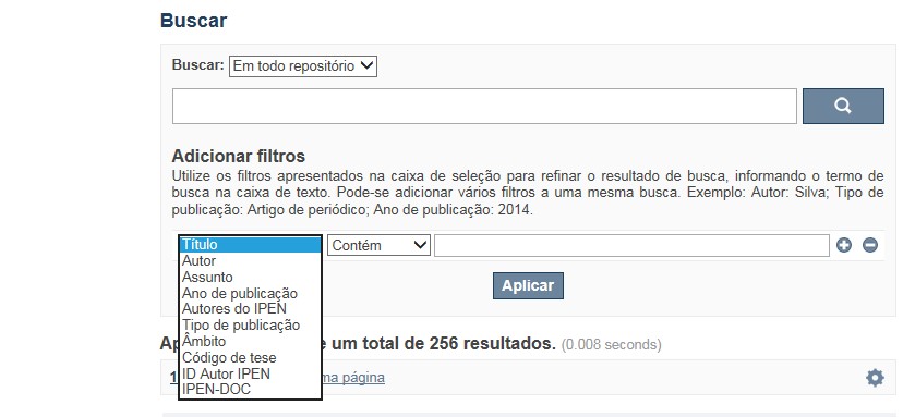
A opção Busca avançada utiliza os conectores da lógica boleana, é o melhor recurso para combinar chaves de busca e obter documentos relevantes à sua pesquisa, utilize os filtros apresentados na caixa de seleção para refinar o resultado de busca. Pode-se adicionar vários filtros a uma mesma busca.
Exemplo:
Buscar os artigos apresentados em um evento internacional de 2015, sobre loss of coolant, do autor Maprelian.
Autor: Maprelian
Título: loss of coolant
Tipo de publicação: Texto completo de evento
Ano de publicação: 2015

✔ Para indexação dos documentos é utilizado o Thesaurus do INIS, especializado na área nuclear e utilizado em todos os países membros da International Atomic Energy Agency – IAEA , por esse motivo, utilize os termos de busca de assunto em inglês; isto não exclui a busca livre por palavras, apenas o resultado pode não ser tão relevante ou pertinente.
✔ 95% do RD apresenta o texto completo do documento com livre acesso, para aqueles que apresentam o ![]() significa que e o documento está sujeito as leis de direitos autorais, solicita-se nesses casos contatar a Biblioteca do IPEN,
bibl@ipen.br
.
significa que e o documento está sujeito as leis de direitos autorais, solicita-se nesses casos contatar a Biblioteca do IPEN,
bibl@ipen.br
.
✔ Ao efetuar a busca por um autor o RD apresentará uma relação de todos os trabalhos depositados no RD. No lado direito da tela são apresentados os coautores com o número de trabalhos produzidos em conjunto bem como os assuntos abordados e os respectivos anos de publicação agrupados.
✔ O RD disponibiliza um quadro estatístico de produtividade, onde é possível visualizar o número dos trabalhos agrupados por tipo de coleção, a medida que estão sendo depositados no RD.
✔ Na página inicial nas referências são sinalizados todos os autores IPEN, ao clicar nesse símbolo ![]() será aberta uma nova página correspondente à aquele autor – trata-se da página do pesquisador.
será aberta uma nova página correspondente à aquele autor – trata-se da página do pesquisador.
✔ Na página do pesquisador, é possível verificar, as variações do nome, a relação de todos os trabalhos com texto completo bem como um quadro resumo numérico; há links para o Currículo Lattes e o Google Acadêmico ( quando esse for informado).
ATENÇÃO!
ESTE TEXTO "AJUDA" ESTÁ SUJEITO A ATUALIZAÇÕES CONSTANTES, A MEDIDA QUE NOVAS FUNCIONALIDADES E RECURSOS DE BUSCA FOREM SENDO DESENVOLVIDOS PELAS EQUIPES DA BIBLIOTECA E DA INFORMÁTICA.
O gerenciamento do Repositório está a cargo da Biblioteca do IPEN. Constam neste RI, até o presente momento 20.950 itens que tanto podem ser artigos de periódicos ou de eventos nacionais e internacionais, dissertações e teses, livros, capítulo de livros e relatórios técnicos. Para participar do RI-IPEN é necessário que pelo menos um dos autores tenha vínculo acadêmico ou funcional com o Instituto. Nesta primeira etapa de funcionamento do RI, a coleta das publicações é realizada periodicamente pela equipe da Biblioteca do IPEN, extraindo os dados das bases internacionais tais como a Web of Science, Scopus, INIS, SciElo além de verificar o Currículo Lattes. O RI-IPEN apresenta também um aspecto inovador no seu funcionamento. Por meio de metadados específicos ele está vinculado ao sistema de gerenciamento das atividades do Plano Diretor anual do IPEN (SIGEPI). Com o objetivo de fornecer dados numéricos para a elaboração dos indicadores da Produção Cientifica Institucional, disponibiliza uma tabela estatística registrando em tempo real a inserção de novos itens. Foi criado um metadado que contém um número único para cada integrante da comunidade científica do IPEN. Esse metadado se transformou em um filtro que ao ser acionado apresenta todos os trabalhos de um determinado autor independente das variáveis na forma de citação do seu nome.
A elaboração do projeto do RI do IPEN foi iniciado em novembro de 2013, colocado em operação interna em julho de 2014 e disponibilizado na Internet em junho de 2015. Utiliza o software livre Dspace, desenvolvido pelo Massachusetts Institute of Technology (MIT). Para descrição dos metadados adota o padrão Dublin Core. É compatível com o Protocolo de Arquivos Abertos (OAI) permitindo interoperabilidade com repositórios de âmbito nacional e internacional.
1. Portaria IPEN-CNEN/SP nº 387, que estabeleceu os princípios que nortearam a criação do RDI, clique aqui.
2. A experiência do Instituto de Pesquisas Energéticas e Nucleares (IPEN-CNEN/SP) na criação de um Repositório Digital Institucional – RDI, clique aqui.
O Repositório Digital do IPEN é um equipamento institucional de acesso aberto, criado com o objetivo de reunir, preservar, disponibilizar e conferir maior visibilidade à Produção Científica publicada pelo Instituto, desde sua criação em 1956.
Operando, inicialmente como uma base de dados referencial o Repositório foi disponibilizado na atual plataforma, em junho de 2015. No Repositório está disponível o acesso ao conteúdo digital de artigos de periódicos, eventos, nacionais e internacionais, livros, capítulos, dissertações, teses e relatórios técnicos.
A elaboração do projeto do RI do IPEN foi iniciado em novembro de 2013, colocado em operação interna em julho de 2014 e disponibilizado na Internet em junho de 2015. Utiliza o software livre Dspace, desenvolvido pelo Massachusetts Institute of Technology (MIT). Para descrição dos metadados adota o padrão Dublin Core. É compatível com o Protocolo de Arquivos Abertos (OAI) permitindo interoperabilidade com repositórios de âmbito nacional e internacional.
O gerenciamento do Repositório está a cargo da Biblioteca do IPEN. Constam neste RI, até o presente momento 20.950 itens que tanto podem ser artigos de periódicos ou de eventos nacionais e internacionais, dissertações e teses, livros, capítulo de livros e relatórios técnicos. Para participar do RI-IPEN é necessário que pelo menos um dos autores tenha vínculo acadêmico ou funcional com o Instituto. Nesta primeira etapa de funcionamento do RI, a coleta das publicações é realizada periodicamente pela equipe da Biblioteca do IPEN, extraindo os dados das bases internacionais tais como a Web of Science, Scopus, INIS, SciElo além de verificar o Currículo Lattes. O RI-IPEN apresenta também um aspecto inovador no seu funcionamento. Por meio de metadados específicos ele está vinculado ao sistema de gerenciamento das atividades do Plano Diretor anual do IPEN (SIGEPI). Com o objetivo de fornecer dados numéricos para a elaboração dos indicadores da Produção Cientifica Institucional, disponibiliza uma tabela estatística registrando em tempo real a inserção de novos itens. Foi criado um metadado que contém um número único para cada integrante da comunidade científica do IPEN. Esse metadado se transformou em um filtro que ao ser acionado apresenta todos os trabalhos de um determinado autor independente das variáveis na forma de citação do seu nome.
