Navegação Periódicos - Artigos por assunto "image processing"
- Página inicial
- →
- IPEN
- →
- Periódicos - Artigos
- →
- Navegação Periódicos - Artigos por assunto
- Sobre
- Perfil Técnico
- Política de funcionamento
- Ajuda
- Apresentação
Navegação Periódicos - Artigos por assunto "image processing"
Itens para a visualização no momento 1-20 de 23
-
. Characteristics of the CsI:Tl scintillator crystal for X-Ray imaging applications. Materials Sciences and Applications, v. 9, n. 2, p. 268-280, 2018. DOI: 10.4236/msa.2018.92018 Abstract: Scintillators are high-density luminescent materials that convert X-rays to visible light. Thallium doped cesium iodide (CsI:Tl) scintillation materials are widely used as converters for X-rays into visible light, with very high conversion efficiency of 64.000 optical photons/MeV. CsI:Tl crystals are commercially available, but, the possibility of developing these crystals into different geometric shapes, meeting the need for coupling the photosensor and reducing cost, makes this material very attractive for scientific research. The objective of this work was to study the feasibility of using radiation sensors, scintillators type, developed for use in imaging systems for X-rays. In this paper, the CsI:Tl scintillator crystal with nominal concentration of the 10−3 M was grown by the vertical Bridgman technique. The imaging performance of CsI:Tl scintillator was studied as a function of the design type and thickness, since it interferes with the light scattering and, hence, the detection efficiency plus final image resolution. The result of the diffraction X-ray analysis in the grown crystals was consistent with the pattern of a face-centered cubic (fcc) crystal structure. Slices 25 × 2 × 3 mm3 (length, thickness, height) of the crystal and mini crystals of 1 × 2 × 3 mm3 (length, thickness, height) were used for comparison in the imaging systems for X-rays. With these crystals scintillators, images of undesirable elements, such as metals in food packaging, were obtained. One-dimensional array of photodiodes and the photosensor CCD (Coupled Charge Device) component were used. In order to determine the ideal thickness of the slices of the scintillator crystal CsI:Tl, Monte Carlo method was used.
Palavras-Chave: cesium iodides; doped materials; thallium ions; crystal growth; phosphors; image processing; photodiodes; charge-coupled devices
. Characteristics of the CsI:Tl scintillator crystal for X-Ray imaging applications. Materials Sciences and Applications, v. 9, n. 2, p. 268-280, 2018. DOI: 10.4236/msa.2018.92018. Disponível em: http://repositorio.ipen.br/handle/123456789/28986. Acesso em: $DATA.Como referenciar este itemEsta referência é gerada automaticamente de acordo com as normas do estilo IPEN/SP (ABNT NBR 6023) e recomenda-se uma verificação final e ajustes caso necessário.
-
. Comparison of TG-43 and TG-186 in breast irradiation using a low energy electronic brachytherapy source. Medical Physics, v. 41, n. 6, p. 061701-1 - 061701-12, 2014.
Palavras-Chave: animal tissues; brachytherapy; computerized tomography; dosimetry; image processing; mammary glands; monte carlo method; patients; radiation dose distributions; radiation doses; recommendations; skin
. Comparison of TG-43 and TG-186 in breast irradiation using a low energy electronic brachytherapy source. Medical Physics, v. 41, n. 6, p. 061701-1 - 061701-12, 2014. Disponível em: http://repositorio.ipen.br/handle/123456789/8940. Acesso em: $DATA.Como referenciar este itemEsta referência é gerada automaticamente de acordo com as normas do estilo IPEN/SP (ABNT NBR 6023) e recomenda-se uma verificação final e ajustes caso necessário.
-
. Determination of dental decay rates with optical coherence tomography. Laser Physics Letters, v. 6, n. 12, p. 896-900, 2009.
Palavras-Chave: image processing; tomography; optical properties; teeth; animal tissues; therapeutic uses; demineralization; lasers
. Determination of dental decay rates with optical coherence tomography. Laser Physics Letters, v. 6, n. 12, p. 896-900, 2009. Disponível em: http://repositorio.ipen.br/handle/123456789/4885. Acesso em: $DATA.Como referenciar este itemEsta referência é gerada automaticamente de acordo com as normas do estilo IPEN/SP (ABNT NBR 6023) e recomenda-se uma verificação final e ajustes caso necessário.
-
. Development and characterisation of a radiation scanning system for small organ studies. Revista Brasileira de Pesquisa e Desenvolvimento, v. 5, n. 1, p. 13-18, 2003.
Palavras-Chave: image processing; organs; thyroid; images; radiation detection; image scanners; iodine 131; technetium 99; diagnosis; diagnostic techniques
. Development and characterisation of a radiation scanning system for small organ studies. Revista Brasileira de Pesquisa e Desenvolvimento, v. 5, n. 1, p. 13-18, 2003. Disponível em: http://repositorio.ipen.br/handle/123456789/5868. Acesso em: $DATA.Como referenciar este itemEsta referência é gerada automaticamente de acordo com as normas do estilo IPEN/SP (ABNT NBR 6023) e recomenda-se uma verificação final e ajustes caso necessário.
-
. Development of ZJU high-spectral-resolution lidar for aerosol and cloud: extinction retrieval. Remote Sensing, v. 12, n. 18, p. 1-18, 2020. DOI: 10.3390/rs12183047 Abstract: The retrieval of the extinction coefficients of aerosols and clouds without assumptions is the most important advantage of the high-spectral-resolution lidar (HSRL). The standard method to retrieve the extinction coefficient from HSRL signals depends heavily on the signal-to-noise ratio (SNR). In this work, an iterative image reconstruction (IIR) method is proposed for the retrieval of the aerosol extinction coefficient based on HSRL data, this proposed method manages to minimize the difference between the reconstructed and raw signals based on reasonable estimates of the lidar ratio. To avoid the ill-posed solution, a regularization method is adopted to reconstruct the lidar signals in the IIR method. The results from Monte-Carlo (MC) simulations applying both standard and IIR methods are compared and these comparisons demonstrate that the extinction coefficient and the lidar ratio retrieved by the IIR method have smaller root mean square error (RMSE) and relative bias values than the standard method. A case study of measurements made by Zhejiang University (ZJU) HSRL is presented, and their results show that the IIR method not only obtains a finer structure of the aerosol layer under the condition of low SNR, but it is also able to retrieve more reasonable values of the lidar ratio.
Palavras-Chave: optical radar; aerosols; monte carlo method; signal-to-noise ratio; iterative methods; image processing; climatic change; scattering
. Development of ZJU high-spectral-resolution lidar for aerosol and cloud: extinction retrieval. Remote Sensing, v. 12, n. 18, p. 1-18, 2020. DOI: 10.3390/rs12183047. Disponível em: http://200.136.52.105/handle/123456789/31652. Acesso em: $DATA.Como referenciar este itemEsta referência é gerada automaticamente de acordo com as normas do estilo IPEN/SP (ABNT NBR 6023) e recomenda-se uma verificação final e ajustes caso necessário.
-
. Digital image processing for biocompatibility studies of clinical implant materials. Artificial Organs, v. 27, n. 5, p. 444-446, 2003.
Palavras-Chave: image processing; computers; scanning electron microscopy; biological materials; blood platelets; polymers; compatibility; prostheses; cardiovascular system
. Digital image processing for biocompatibility studies of clinical implant materials. Artificial Organs, v. 27, n. 5, p. 444-446, 2003. Disponível em: http://repositorio.ipen.br/handle/123456789/7454. Acesso em: $DATA.Como referenciar este itemEsta referência é gerada automaticamente de acordo com as normas do estilo IPEN/SP (ABNT NBR 6023) e recomenda-se uma verificação final e ajustes caso necessário.
-
. Dose-image quality study in digital chest radiography using Monte Carlo simulation. Applied Radiation and Isotopes, v. 66, n. 9, p. 1213-1217, 2008.
Palavras-Chave: chest; biomedical radiography; radiation dose; image processing; quality assurance; simulation; monte carlo method; m codes; digital systems
. Dose-image quality study in digital chest radiography using Monte Carlo simulation. Applied Radiation and Isotopes, v. 66, n. 9, p. 1213-1217, 2008. Disponível em: http://repositorio.ipen.br/handle/123456789/7975. Acesso em: $DATA.Como referenciar este itemEsta referência é gerada automaticamente de acordo com as normas do estilo IPEN/SP (ABNT NBR 6023) e recomenda-se uma verificação final e ajustes caso necessário.
-
. Emission and transmission tomography system applied to analyze industrial process inside chemical reactors. Nuclear Instruments and Methods in Physics Research Section A: Accelerators, Spectrometers, Detectors and Associated Equipment, v. 954, p. 1-3, 2020. DOI: 10.1016/j.nima.2019.01.073 Abstract: The tomography techniques are widely used in many industries, such as: chemical, food, pharmaceutical and oil sectors. In the industries the tomography is used to diagnose the state of the machines of production and also in the control of quality of the produced objects. A portable tomography system known as instant-non-scanning type, a similar version of the fourth generation CT, was developed in this work. It is capable to obtain measurements in real time conditions without interrupting the operation of the industrial production and it is useful in the quality control of the means of production and the objects produced. This paper describes an innovative hybrid industrial tomographic system, i.e., simultaneous data from the emission of an internal radioactive source introduced inside to the object (67Ga citrate) and tomographic transmission using five sources of 137Cs positioned externally to the object which are distributed at the vertices of a pentagon. The tomographic system described here is useful for studying dynamic chemical phenomena, associated or not with multiphase systems commonly found in chemical reactors and distillation columns.
Palavras-Chave: cesium 137; diagnostic techniques; tomography; computerized tomography; image processing; nai detectors; citrates; gallium 67; external irradiation; trace amounts
. Emission and transmission tomography system applied to analyze industrial process inside chemical reactors. Nuclear Instruments and Methods in Physics Research Section A: Accelerators, Spectrometers, Detectors and Associated Equipment, v. 954, p. 1-3, 2020. DOI: 10.1016/j.nima.2019.01.073. Disponível em: http://repositorio.ipen.br/handle/123456789/31101. Acesso em: $DATA.Como referenciar este itemEsta referência é gerada automaticamente de acordo com as normas do estilo IPEN/SP (ABNT NBR 6023) e recomenda-se uma verificação final e ajustes caso necessário.
-
. Environmental tobacco smoke during the early postnatal period of mice interferes with brain 18F-FDG uptake from infancy to early adulthood: a longitudinal study. Frontiers in Neuroscience, v. 14, p. 1-11, 2020. DOI: 10.3389/fnins.2020.00005 Abstract: Exposure to environmental tobacco smoke (ETS) is associated with high morbidity and mortality, mainly in childhood. Our aim was to evaluate the effects of postnatal ETS exposure in the brain 2-deoxy-2-[18F]-fluoro-D-glucose (18F-FDG) uptake of mice by positron emission tomography (PET) neuroimaging in a longitudinal study. C57BL/6J mice were exposed to ETS that was generated from 3R4F cigarettes from postnatal day 3 (P3) to P14. PET analyses were performed in male and female mice during infancy (P15), adolescence (P35), and adulthood (P65). We observed that ETS exposure decreased 18F-FDG uptake in the whole brain, both left and right hemispheres, and frontal cortex in both male and female infant mice, while female infant mice exposed to ETS showed decreased 18F-FDG uptake in the cerebellum. In addition, all mice showed reduced 18F-FDG uptake in infancy, compared to adulthood in all analyzed VOIs. In adulthood, ETS exposure during the early postnatal period decreased brain 18FFDG uptake in adult male mice in the cortex, striatum, hippocampus, cingulate cortex, and thalamus when compared to control group. ETS induced an increase in 18F-FDG uptake in adult female mice when compared to control group in the brainstem and cingulate cortex. Moreover, male ETS-exposed animals showed decreased 18F-FDG uptake when compared to female ETS-exposed in the whole brain, brainstem, cortex, left amygdala, striatum, hippocampus, cingulate cortex, basal forebrain and septum, thalamus, hypothalamus, and midbrain. The present study shows that several brain regions are vulnerable to ETS exposure during the early postnatal period and these effects on 18F-FDG uptake are observed even a long time after the last exposure. This study corroborates our previous findings, strengthening the idea that exposure to tobacco smoke in a critical period interferes with brain development of mice from late infancy to early adulthood.
Palavras-Chave: tobacco; tobacco smokes; nicotiana; environment; image processing; positron computed tomography; uptake; fluorodeoxyglucose; mice; glucose; longitudinal momentum; brain; children; nerve cells
. Environmental tobacco smoke during the early postnatal period of mice interferes with brain 18F-FDG uptake from infancy to early adulthood: a longitudinal study. Frontiers in Neuroscience, v. 14, p. 1-11, 2020. DOI: 10.3389/fnins.2020.00005. Disponível em: http://repositorio.ipen.br/handle/123456789/31363. Acesso em: $DATA.Como referenciar este itemEsta referência é gerada automaticamente de acordo com as normas do estilo IPEN/SP (ABNT NBR 6023) e recomenda-se uma verificação final e ajustes caso necessário.
-
. Image quality analysis of an industrial instant non-scanning tomography system with different collimators. Brazilian Journal of Radiation Sciences, v. 7, n. 2A, p. 1-14, 2019. DOI: 10.15392/bjrs.v7i2A.561 Abstract: An instant non-scanning tomography system is being developed at the IPEN. This type of tomography system, due to its configurations, provides a low spatial resolution; however, it presents a high temporal resolution as an advantage. In this sense, it has high applicability in industrial processes involving dynamic multiphase processes, like distillation columns. Thereby, to obtain the best conditions of image acquisition, this tomography system comprising different collimators was simulated with Monte Carlo using the MCNP4C.The image quality was evaluated with Matlab® 2013b analyzing the contrast to noise ratio (CNR), root mean square ratio (RMSE), signal to noise ratio (SNR) and the spatial resolution by the Modulation Transfer Function (MTF(f)), to identify which collimator fits better to the tomography in development. It was simulated three situations; (i) with no collimator; (ii) ø 25x 50 mm2 cylindrical collimator with a septum of ø5.0 x 50 mm2; (iii) ø25 x 50 mm2 cylindrical collimator with a slit septum of 24 x 5.0 x 50 mm3. RMSE values for no collimator presented better results. CNR showed that no collimator and slit collimator reaches the same CNR values, but no collimator decreases more than the slit collimator as the number of iteration rises. The hole collimator reaches a higher CNR value, however decreases more than the others. The spatial resolution with no collimator and slit collimator were around 31.9 mm, and for the hole collimator was around 20 mm.
Palavras-Chave: collimators; tomography; computerized tomography; computerized simulation; monte carlo method; quality assurance; quality management; image processing; nai detectors; signal-to-noise ratio; radiation detectors; radiation detectors; industrial plants; brazilian cnen
. Image quality analysis of an industrial instant non-scanning tomography system with different collimators. Brazilian Journal of Radiation Sciences, v. 7, n. 2A, p. 1-14, 2019. DOI: 10.15392/bjrs.v7i2A.561. Disponível em: http://repositorio.ipen.br/handle/123456789/29991. Acesso em: $DATA.Como referenciar este itemEsta referência é gerada automaticamente de acordo com as normas do estilo IPEN/SP (ABNT NBR 6023) e recomenda-se uma verificação final e ajustes caso necessário.
-
. A medical image-based graphical platform-Features, applications and relevance for brachytherapy. Brachytherapy, v. 13, n. 6, p. 632-639, 2014.
Palavras-Chave: brachytherapy; images; image processing; monte carlo method; radiation doses; calculation methods; algorithms; simulation; computer codes
. A medical image-based graphical platform-Features, applications and relevance for brachytherapy. Brachytherapy, v. 13, n. 6, p. 632-639, 2014. Disponível em: http://repositorio.ipen.br/handle/123456789/23185. Acesso em: $DATA.Como referenciar este itemEsta referência é gerada automaticamente de acordo com as normas do estilo IPEN/SP (ABNT NBR 6023) e recomenda-se uma verificação final e ajustes caso necessário.
-
. Methodological analysis of gamma tomography system for large random packed columns. Applied Radiation and Isotopes, v. 68, p. 658-661, 2010.
Palavras-Chave: computerized tomography; gamma radiation; porosity; image processing
. Methodological analysis of gamma tomography system for large random packed columns. Applied Radiation and Isotopes, v. 68, p. 658-661, 2010. Disponível em: http://repositorio.ipen.br/handle/123456789/8066. Acesso em: $DATA.Como referenciar este itemEsta referência é gerada automaticamente de acordo com as normas do estilo IPEN/SP (ABNT NBR 6023) e recomenda-se uma verificação final e ajustes caso necessário.
-
. Nanoradiopharmaceuticals for breast cancer imaging: development, characterization, and imaging in inducted animals. OncoTargets and Therapy, v. 9, p. 5847-5854, 2016. DOI: 10.2147/OTT.S110787 Abstract: Monoclonal antibodies as polymeric nanoparticles are quite interesting and endow this new drug category with many advantages, especially by reducing the number of adverse reactions and, in the case of radiopharmaceuticals, also reducing the amount of radiation (dose) administered to the patient. In this study, a nanoradiopharmaceutical was developed using polylactic acid (PLA)/polyvinyl alcohol (PVA)/montmorillonite (MMT)/trastuzumab nanoparticles labeled with technetium-99m (99mTc) for breast cancer imaging. In order to confirm the nanoparticle formation, atomic force microscopy and dynamic light scattering were performed. Cytotoxicity of the nanoparticle and biodistribution with 99mTc in healthy and inducted animals were also measured. The results from atomic force microscopy showed that the nanoparticles were spherical, with a size range of ~200–500 nm. The dynamic light scattering analysis demonstrated that over 90% of the nanoparticles produced had a size of 287 nm with a zeta potential of -14,6 mV. The cytotoxicity results demonstrated that the nanoparticles were capable of reaching breast cancer cells. The biodistribution data demonstrated that the PLA/PVA/MMT/trastuzumab nanoparticles labeled with 99mTc have great renal clearance and also a high uptake by the lesion, as ~45% of the PLA/PVA/MMT/trastuzumab nanoparticles injected were taken up by the lesion. The data support PLA/PVA/MMT/trastuzumab labeled with 99mTc nanoparticles as nanoradiopharmaceuticals for breast cancer imaging.
Palavras-Chave: medicine; neoplasms; radiopharmaceuticals; nanotechnology; mammary glands; molecular structure; image processing; nanostructures; diagnosis; nuclear medicine
. Nanoradiopharmaceuticals for breast cancer imaging: development, characterization, and imaging in inducted animals. OncoTargets and Therapy, v. 9, p. 5847-5854, 2016. DOI: 10.2147/OTT.S110787. Disponível em: http://repositorio.ipen.br/handle/123456789/26786. Acesso em: $DATA.Como referenciar este itemEsta referência é gerada automaticamente de acordo com as normas do estilo IPEN/SP (ABNT NBR 6023) e recomenda-se uma verificação final e ajustes caso necessário.
-
. Octreotide nanoparticles showed affinity for in vivo MIA Paca-2 inducted pancreas ductal adenocarcinoma mimicking pancreatic polypeptide-secreting tumor of the distal pancreas (PPoma). Pharmaceutical Research, v. 36, n. 10, p. 1-8, 2019. DOI: 10.1007/s11095-019-2678-4 Abstract: Purpose Pancreatic Polypeptide-secreting tumor of the distal pancreas (PPoma) is a rare, difficult and indolent type of cancer with a survival rate of 5-year in only 10% of all cases. The PPoma is classified as a neuroendocrine tumor (NET) not functioning that overexpresses SSTR 2 (somatostatin receptor subtype 2). Thus, in order to improve the diagnosis of this type of tumor, we developed nanoparticulate drug carriers based on poly-lactic acid (PLA) polymer loaded with octreotide and radiolabeled with Technetium-99 m (99mTc). Methods PLA/PVA octreotide nanoparticles were developed by double-emulsion technique. These nanoparticles were characterized by Atomic Force Microscopy (AFM) and Dynamic Light Scattering (DLS) and radiolabeled with 99mTc by the direct via forming 99mTc-PLA/PVA octreotide nanoparticles. The safety of these nanosystems was evaluated by the MTT cell toxicity assay and their in vivo biodistribution was evaluated in xenografted inducted animals. Results The results showed that a 189 nm sized nanoparticle were formed with a PDI of 0,097, corroborating the monodispersive behavior. These nanoparticles were successfully radiolabeled with 99mTc showing uptake by the inducted tumor. The MTT assay corroborated the safety of the nanosystem for the cells. Conclusion The results support the use of this nanosystem (99mTc-PLA/PVA octreotide nanoparticles) as imaging agent for PPoma.
Palavras-Chave: nanoparticles; radiopharmaceuticals; pancreas; carcinomas; images; image processing; light scattering; tumor cells; cell cultures
. Octreotide nanoparticles showed affinity for in vivo MIA Paca-2 inducted pancreas ductal adenocarcinoma mimicking pancreatic polypeptide-secreting tumor of the distal pancreas (PPoma). Pharmaceutical Research, v. 36, n. 10, p. 1-8, 2019. DOI: 10.1007/s11095-019-2678-4. Disponível em: http://repositorio.ipen.br/handle/123456789/30459. Acesso em: $DATA.Como referenciar este itemEsta referência é gerada automaticamente de acordo com as normas do estilo IPEN/SP (ABNT NBR 6023) e recomenda-se uma verificação final e ajustes caso necessário.
-
. Phase quantification in UAlx-Al dispersion targets for Mo-99 production. Journal of Nuclear Materials, v. 509, p. 465-477, 2018. DOI: 10.1016/j.jnucmat.2018.07.029 Abstract: Uranium aluminide (UAlx) is a mixture of three distinct intermetallic compounds comprised of UAl2, UAl3 and UAl4, where the “x” is used to denote a mixture of those phases. Usually UAlx is formed during the target fabrication process by means of a solid state reaction between the uranium aluminide and aluminum. Quantitative techniques such as image analysis and X-ray diffraction using the Rietveld method were compared for their applicability in the determination of the UAl2, UAl3 and UAl4 concentrations, both in the UAl2 primary ingot and in the UAlx-Al dispersion. The UAlx composition was quantified in all stages of the target manufacturing. The image analysis method was shown to be useful for UAlx phase quantification in the primary UAl2 ingot, but was not applicable in the case of UAlx-Al dispersions. The X-ray diffraction method allowed the quantification of the existing UAlx phases in both the primary ingot and UAlx-Al dispersions. Possible sources of error are discussed. The method of quantification based on X-ray diffraction was shown to be appropriate to monitor the evolution of UAlx phases during the manufacturing process.
Palavras-Chave: diffraction methods; dispersions; image processing; mapping; molybdenum 99; x-ray diffraction
. Phase quantification in UAlx-Al dispersion targets for Mo-99 production. Journal of Nuclear Materials, v. 509, p. 465-477, 2018. DOI: 10.1016/j.jnucmat.2018.07.029. Disponível em: http://repositorio.ipen.br/handle/123456789/29293. Acesso em: $DATA.Como referenciar este itemEsta referência é gerada automaticamente de acordo com as normas do estilo IPEN/SP (ABNT NBR 6023) e recomenda-se uma verificação final e ajustes caso necessário.
-
. Pixel-position-based lossless image compression algorithm. International Journal of Innovative Studies in Sciences and Engineering Technology, v. 5, n. 12, p. 21-30, 2019. Abstract: In this paper we present a novel lossless image compression method that is very simple and fast. The method uses linear prediction followed by arithmetic coding. Different prediction functions are used to estimate the intensity of image pixels. Two variants of the prediction algorithm are presented. One variant uses two different prediction functions and the other uses three different prediction functions. The position of the pixel in the image determines which prediction function is used. The method can be applied for images of any size and of high bit-depths. Standard images available in the literature are used to test the method. The compression ratios obtained with the proposed method are compared with the compression ratios obtained with the JPEG-LS and JPEG2000 methods and the results are satisfactory.
Palavras-Chave: images; image processing; compression; algorithms; computer graphics; computer codes; image converters; standardization; tomography; x-ray equipment; ultrasonography; oil spills; biomedical radiography
. Pixel-position-based lossless image compression algorithm. International Journal of Innovative Studies in Sciences and Engineering Technology, v. 5, n. 12, p. 21-30, 2019. Disponível em: http://repositorio.ipen.br/handle/123456789/30846. Acesso em: $DATA.Como referenciar este itemEsta referência é gerada automaticamente de acordo com as normas do estilo IPEN/SP (ABNT NBR 6023) e recomenda-se uma verificação final e ajustes caso necessário.
-
. Quantification of microstructural features in gadolinia-doped ceria containing co-additives by digital image analysis. Journal of the European Ceramic Society, v. 31, n. 8, p. 1431-1438, 2011.
Palavras-Chave: gadolinium oxides; doped materials; cerium oxides; powders; synthesis; microstructure; porosity; image processing; digital systems
. Quantification of microstructural features in gadolinia-doped ceria containing co-additives by digital image analysis. Journal of the European Ceramic Society, v. 31, n. 8, p. 1431-1438, 2011. Disponível em: http://repositorio.ipen.br/handle/123456789/4500. Acesso em: $DATA.Como referenciar este itemEsta referência é gerada automaticamente de acordo com as normas do estilo IPEN/SP (ABNT NBR 6023) e recomenda-se uma verificação final e ajustes caso necessário.
-
. Real-time neutron radiography at the IEA-R1m nuclear research reactor. Brazilian Journal of Physics, v. 33, n. 2, special issue, p. 282-285, 2003.
Palavras-Chave: neutron radiography; real time systems; image intensifiers; images; image processing; sensitivity; iear-1 reactor
. Real-time neutron radiography at the IEA-R1m nuclear research reactor. Brazilian Journal of Physics, v. 33, n. 2, p. 282-285, 2003. special issue. Disponível em: http://repositorio.ipen.br/handle/123456789/5818. Acesso em: $DATA.Como referenciar este itemEsta referência é gerada automaticamente de acordo com as normas do estilo IPEN/SP (ABNT NBR 6023) e recomenda-se uma verificação final e ajustes caso necessário.
-
. Results of process equipment imaging by tomographic gamma scan. Chemical Engineering Transactions, v. 69, p. 649-654, 2018. DOI: 10.3303/CET1869109 Abstract: Gamma scanning is a nuclear inspection technique widely used for troubleshooting industrial process equipments in refineries and petrochemicals. A radiation source and detector move vertically along the height of the vessel and the radiation readings are used to plot the graph of density profile. In recent years, many improvements have been added to this technique, but essentially the result of gamma scanning still consists of a 1-D density plot. Developed in 2012, the tomographic gamma scan uses image reconstruction techniques to show the result of gamma scan as a 2-D density distribution image. In this work, we present the current status of the technology and the results obtained by applying the technique to real operating process equipment. The results present images that match well with the 1-D density plots obtained by the conventional gamma scan and reveal process and operational problems that has never been visualized before with the traditional technique. The tomographic gamma scan can be immediately applied in practice with low financial investment.
Palavras-Chave: image processing; tomography; gamma radiation; equipment; gamma fuel scanning; nondestructive testing; irradiation; radiation sources; electron scanning; gamma radiography; tomography; gamma radiation; distillation; gamma sources; irradiation
. Results of process equipment imaging by tomographic gamma scan. Chemical Engineering Transactions, v. 69, p. 649-654, 2018. DOI: 10.3303/CET1869109. Disponível em: http://repositorio.ipen.br/handle/123456789/29424. Acesso em: $DATA.Como referenciar este itemEsta referência é gerada automaticamente de acordo com as normas do estilo IPEN/SP (ABNT NBR 6023) e recomenda-se uma verificação final e ajustes caso necessário.
-
. Study of cracking in concrete by neutron radiography. Applied Radiation and Isotopes, v. 48, n. 3, p. 339-344, 1997.
Palavras-Chave: concretes; cracks; neutron radiography; image processing
. Study of cracking in concrete by neutron radiography. Applied Radiation and Isotopes, v. 48, n. 3, p. 339-344, 1997. Disponível em: http://repositorio.ipen.br/handle/123456789/6409. Acesso em: $DATA.Como referenciar este itemEsta referência é gerada automaticamente de acordo com as normas do estilo IPEN/SP (ABNT NBR 6023) e recomenda-se uma verificação final e ajustes caso necessário.
Itens para a visualização no momento 1-20 de 23
Buscar no repositório
Navegar
Minha conta
Visualizar
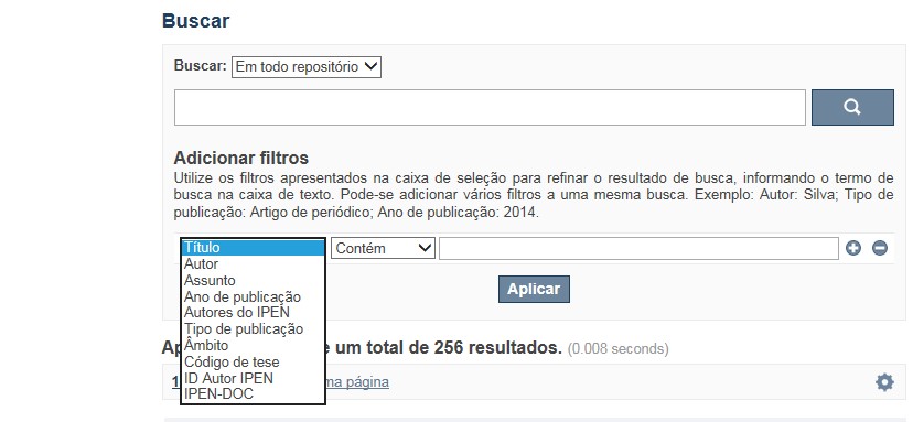
A pesquisa no RD utiliza os recursos de busca da maioria das bases de dados. No entanto algumas dicas podem auxiliar para obter um resultado mais pertinente.
✔ É possível efetuar a busca de um autor ou um termo em todo o RD, por meio do Buscar no Repositório , isto é, o termo solicitado será localizado em qualquer campo do RD. No entanto esse tipo de pesquisa não é recomendada a não ser que se deseje um resultado amplo e generalizado.
✔ A pesquisa apresentará melhor resultado selecionando um dos filtros disponíveis em Navegar
✔ Os filtros disponíveis em Navegar tais como: Coleções, Ano de publicação, Títulos, Assuntos, Autores, Revista, Tipo de publicação são autoexplicativos. O filtro, Autores IPEN apresenta uma relação com os autores vinculados ao IPEN; o ID Autor IPEN diz respeito ao número único de identificação de cada autor constante no RD e sob o qual estão agrupados todos os seus trabalhos independente das variáveis do seu nome; Tipo de acesso diz respeito à acessibilidade do documento, isto é , sujeito as leis de direitos autorais, ID RT apresenta a relação dos relatórios técnicos, restritos para consulta das comunidades indicadas.

A opção Busca avançada utiliza os conectores da lógica boleana, é o melhor recurso para combinar chaves de busca e obter documentos relevantes à sua pesquisa, utilize os filtros apresentados na caixa de seleção para refinar o resultado de busca. Pode-se adicionar vários filtros a uma mesma busca.
Exemplo:
Buscar os artigos apresentados em um evento internacional de 2015, sobre loss of coolant, do autor Maprelian.
Autor: Maprelian
Título: loss of coolant
Tipo de publicação: Texto completo de evento
Ano de publicação: 2015

✔ Para indexação dos documentos é utilizado o Thesaurus do INIS, especializado na área nuclear e utilizado em todos os países membros da International Atomic Energy Agency – IAEA , por esse motivo, utilize os termos de busca de assunto em inglês; isto não exclui a busca livre por palavras, apenas o resultado pode não ser tão relevante ou pertinente.
✔ 95% do RD apresenta o texto completo do documento com livre acesso, para aqueles que apresentam o ![]() significa que e o documento está sujeito as leis de direitos autorais, solicita-se nesses casos contatar a Biblioteca do IPEN,
bibl@ipen.br
.
significa que e o documento está sujeito as leis de direitos autorais, solicita-se nesses casos contatar a Biblioteca do IPEN,
bibl@ipen.br
.
✔ Ao efetuar a busca por um autor o RD apresentará uma relação de todos os trabalhos depositados no RD. No lado direito da tela são apresentados os coautores com o número de trabalhos produzidos em conjunto bem como os assuntos abordados e os respectivos anos de publicação agrupados.
✔ O RD disponibiliza um quadro estatístico de produtividade, onde é possível visualizar o número dos trabalhos agrupados por tipo de coleção, a medida que estão sendo depositados no RD.
✔ Na página inicial nas referências são sinalizados todos os autores IPEN, ao clicar nesse símbolo ![]() será aberta uma nova página correspondente à aquele autor – trata-se da página do pesquisador.
será aberta uma nova página correspondente à aquele autor – trata-se da página do pesquisador.
✔ Na página do pesquisador, é possível verificar, as variações do nome, a relação de todos os trabalhos com texto completo bem como um quadro resumo numérico; há links para o Currículo Lattes e o Google Acadêmico ( quando esse for informado).
ATENÇÃO!
ESTE TEXTO "AJUDA" ESTÁ SUJEITO A ATUALIZAÇÕES CONSTANTES, A MEDIDA QUE NOVAS FUNCIONALIDADES E RECURSOS DE BUSCA FOREM SENDO DESENVOLVIDOS PELAS EQUIPES DA BIBLIOTECA E DA INFORMÁTICA.
O gerenciamento do Repositório está a cargo da Biblioteca do IPEN. Constam neste RI, até o presente momento 20.950 itens que tanto podem ser artigos de periódicos ou de eventos nacionais e internacionais, dissertações e teses, livros, capítulo de livros e relatórios técnicos. Para participar do RI-IPEN é necessário que pelo menos um dos autores tenha vínculo acadêmico ou funcional com o Instituto. Nesta primeira etapa de funcionamento do RI, a coleta das publicações é realizada periodicamente pela equipe da Biblioteca do IPEN, extraindo os dados das bases internacionais tais como a Web of Science, Scopus, INIS, SciElo além de verificar o Currículo Lattes. O RI-IPEN apresenta também um aspecto inovador no seu funcionamento. Por meio de metadados específicos ele está vinculado ao sistema de gerenciamento das atividades do Plano Diretor anual do IPEN (SIGEPI). Com o objetivo de fornecer dados numéricos para a elaboração dos indicadores da Produção Cientifica Institucional, disponibiliza uma tabela estatística registrando em tempo real a inserção de novos itens. Foi criado um metadado que contém um número único para cada integrante da comunidade científica do IPEN. Esse metadado se transformou em um filtro que ao ser acionado apresenta todos os trabalhos de um determinado autor independente das variáveis na forma de citação do seu nome.
A elaboração do projeto do RI do IPEN foi iniciado em novembro de 2013, colocado em operação interna em julho de 2014 e disponibilizado na Internet em junho de 2015. Utiliza o software livre Dspace, desenvolvido pelo Massachusetts Institute of Technology (MIT). Para descrição dos metadados adota o padrão Dublin Core. É compatível com o Protocolo de Arquivos Abertos (OAI) permitindo interoperabilidade com repositórios de âmbito nacional e internacional.
1. Portaria IPEN-CNEN/SP nº 387, que estabeleceu os princípios que nortearam a criação do RDI, clique aqui.
2. A experiência do Instituto de Pesquisas Energéticas e Nucleares (IPEN-CNEN/SP) na criação de um Repositório Digital Institucional – RDI, clique aqui.
O Repositório Digital do IPEN é um equipamento institucional de acesso aberto, criado com o objetivo de reunir, preservar, disponibilizar e conferir maior visibilidade à Produção Científica publicada pelo Instituto, desde sua criação em 1956.
Operando, inicialmente como uma base de dados referencial o Repositório foi disponibilizado na atual plataforma, em junho de 2015. No Repositório está disponível o acesso ao conteúdo digital de artigos de periódicos, eventos, nacionais e internacionais, livros, capítulos, dissertações, teses e relatórios técnicos.
A elaboração do projeto do RI do IPEN foi iniciado em novembro de 2013, colocado em operação interna em julho de 2014 e disponibilizado na Internet em junho de 2015. Utiliza o software livre Dspace, desenvolvido pelo Massachusetts Institute of Technology (MIT). Para descrição dos metadados adota o padrão Dublin Core. É compatível com o Protocolo de Arquivos Abertos (OAI) permitindo interoperabilidade com repositórios de âmbito nacional e internacional.
O gerenciamento do Repositório está a cargo da Biblioteca do IPEN. Constam neste RI, até o presente momento 20.950 itens que tanto podem ser artigos de periódicos ou de eventos nacionais e internacionais, dissertações e teses, livros, capítulo de livros e relatórios técnicos. Para participar do RI-IPEN é necessário que pelo menos um dos autores tenha vínculo acadêmico ou funcional com o Instituto. Nesta primeira etapa de funcionamento do RI, a coleta das publicações é realizada periodicamente pela equipe da Biblioteca do IPEN, extraindo os dados das bases internacionais tais como a Web of Science, Scopus, INIS, SciElo além de verificar o Currículo Lattes. O RI-IPEN apresenta também um aspecto inovador no seu funcionamento. Por meio de metadados específicos ele está vinculado ao sistema de gerenciamento das atividades do Plano Diretor anual do IPEN (SIGEPI). Com o objetivo de fornecer dados numéricos para a elaboração dos indicadores da Produção Cientifica Institucional, disponibiliza uma tabela estatística registrando em tempo real a inserção de novos itens. Foi criado um metadado que contém um número único para cada integrante da comunidade científica do IPEN. Esse metadado se transformou em um filtro que ao ser acionado apresenta todos os trabalhos de um determinado autor independente das variáveis na forma de citação do seu nome.
