Navegação Periódicos - Artigos por assunto "images"
- Página inicial
- →
- IPEN
- →
- Periódicos - Artigos
- →
- Navegação Periódicos - Artigos por assunto
- Sobre
- Perfil Técnico
- Política de funcionamento
- Ajuda
- Apresentação
Navegação Periódicos - Artigos por assunto "images"
Itens para a visualização no momento 1-20 de 43
-
. Analise de fatores para adoção da tecnologia nuclear no diagnóstico e tratamento de doenças crônicas / Factor analysis for the adoption of nuclear technology in diagnosis and treatment of chronic diseases. Revista Einstein, v. 10, n. 1, p. 62-66, 2012. DOI: 10.1590/s1679-45082012000100013
Palavras-Chave: diseases; diagnostic techniques; images; therapeutic uses; nuclear medicine; radioisotopes; health services
. Analise de fatores para adoção da tecnologia nuclear no diagnóstico e tratamento de doenças crônicas. Revista Einstein, v. 10, n. 1, p. 62-66, 2012. DOI: 10.1590/s1679-45082012000100013. Disponível em: http://repositorio.ipen.br/handle/123456789/4127. Acesso em: $DATA.Como referenciar este itemEsta referência é gerada automaticamente de acordo com as normas do estilo IPEN/SP (ABNT NBR 6023) e recomenda-se uma verificação final e ajustes caso necessário.
-
. AP and PA thorax radiographs: dose evaluation using the FAX phantom. International Journal of Low Radiation, v. 5, n. 3, p. 237-255, 2008.
Palavras-Chave: chest; images; monte carlo method; simulation; females; adults; phantoms; radiation doses
. AP and PA thorax radiographs: dose evaluation using the FAX phantom. International Journal of Low Radiation, v. 5, n. 3, p. 237-255, 2008. Disponível em: http://repositorio.ipen.br/handle/123456789/4904. Acesso em: $DATA.Como referenciar este itemEsta referência é gerada automaticamente de acordo com as normas do estilo IPEN/SP (ABNT NBR 6023) e recomenda-se uma verificação final e ajustes caso necessário.
-
. Assessment of protocols in CT with symmetric and asymmetric beans usingeffective dose and air kerma-area product. Applied Radiation and Isotopes, v. 100, p. 16-20, 2015.
Palavras-Chave: dentistry; radiology; kerma; symmetry; images; cones; computerized tomography; radiation doses; thermoluminescent dosimetry; radiation protection
. Assessment of protocols in CT with symmetric and asymmetric beans usingeffective dose and air kerma-area product. Applied Radiation and Isotopes, v. 100, p. 16-20, 2015. Disponível em: http://repositorio.ipen.br/handle/123456789/23791. Acesso em: $DATA.Como referenciar este itemEsta referência é gerada automaticamente de acordo com as normas do estilo IPEN/SP (ABNT NBR 6023) e recomenda-se uma verificação final e ajustes caso necessário.
-
. Cation distribution of Mn-Zn ferrite nanoparticles using pair distribution function analysis and resonant X-ray scattering. EPL, v. 124, n. 5, p. 56001-p1 - 56001-p5, 2018. DOI: 10.1209/0295-5075/124/56001 Abstract: Mn-Zn ferrite nanoparticles were synthesized by chemical co-precipitation method and analysed using X-ray synchrotron diffraction data. Pair distribution function (PDF) analysis was used to probe the local structure and revealed that the first-neighbour distances of Fe-Fe and Mn-Zn in the 3.0 up to 3.5˚A range are different from the ones usually reported in the literature. For the sample with the best magnetic behaviour, resonant X-ray scattering (RXS) using three energies close to the absorption edges of Mn, Zn and Fe was applied to determine the cation distribution which explained the previous result from PDF analysis.
Palavras-Chave: nanoparticles; distribution functions; scattering; x-ray diffraction; ferrite; zinc compounds; manganese compounds; magnetic resonance; nmr imaging; images
. Cation distribution of Mn-Zn ferrite nanoparticles using pair distribution function analysis and resonant X-ray scattering. EPL, v. 124, n. 5, p. 56001-p1 - 56001-p5, 2018. DOI: 10.1209/0295-5075/124/56001. Disponível em: http://repositorio.ipen.br/handle/123456789/29931. Acesso em: $DATA.Como referenciar este itemEsta referência é gerada automaticamente de acordo com as normas do estilo IPEN/SP (ABNT NBR 6023) e recomenda-se uma verificação final e ajustes caso necessário.
-
. Cell response of calcium phosphate based ceramics, a bone subtitute material. Materials Research, v. 16, n. 4, p. 1052-1057, 2013.
Palavras-Chave: bone tissues; transplants; grafts; biological materials; ceramics; calcium phosphates; scanning electron microscopy; images
. Cell response of calcium phosphate based ceramics, a bone subtitute material. Materials Research, v. 16, n. 4, p. 1052-1057, 2013. Disponível em: http://repositorio.ipen.br/handle/123456789/4014. Acesso em: $DATA.Como referenciar este itemEsta referência é gerada automaticamente de acordo com as normas do estilo IPEN/SP (ABNT NBR 6023) e recomenda-se uma verificação final e ajustes caso necessário.
-
. Characterization and comparative analysis of voids in class II composite resin restorations by optical coherence tomography. Operative Dentistry, v. 45, n. 1, p. 71-79, 2020. DOI: 10.2341/18-290-L Abstract: Purpose: This study aimed to characterize and analyze the number of voids and the percentage of void volume within and between the layers of class II composite restorations made using the bulk fill technique or the incremental technique by optical coherence tomography (OCT). Methods and Materials: Class II cavities (43432 mm) were prepared in 48 human third molars (n=24 restorations per group, two class II cavities per tooth). Teeth were divided into four groups and restored as follows: group 1 (FOB), bulk filled in a single increment using Filtek One Bulk Fill (3M Oral Care); group 2 (FXT), incrementally filled using four oblique layers of Filtek Z350 XT (3M Oral Care); group 3 (FBF+FXT), bulk filled in a single increment using Filtek Bulk Fill Flowable Restorative (3M Oral Care) covered with two oblique layers of Filtek Z350 XT (3M Oral Care), and group 4 (FF+FXT), incrementally filled using Filtek Z350 XT Flow (3M Oral Care) covered with two oblique layers of Filtek Z350 XT (3M Oral Care). After the restorative procedure, specimens were immersed into distilled water and stored in a hot-air oven at 378C. Forty-eight hours later, thermal cycling was conducted (5000 cycles, 58C to 558C). Afterward, OCT was used to detect the existence of voids and to calculate the number of voids and percentage of voids volume within each restoration. Data were submitted to chi-square and Kruskal- Wallis tests (a=0.05). Comparisons were made using the Dunn method. Results: Voids were detected in all groups, ranging from 0.000002 (FBF+FXT and FF+FXT) to 0.32 mm3 (FBF+FXT). FF + FXT presented voids in all of the restorations and had a significantly higher number of voids per restoration when compared to the other groups (p,0.05), but restorations with the presence of voids were significantly higher only when compared to FXT (p,0.05). FBF + FXT presented a significantly higher percentage of voids volume than that of FXT (p,0.05). When comparing restorations made using high-viscosity resin-based composites (FOB and FXT), no significant differences regarding number of voids or percentage of voids volume were detected (p 0.05). Conclusions: The use of flowable resin-based composites can result in an increased number of voids and percentage of voids volume in restorations, and this appears to be more related to voids present inside the syringe of the material than to the use of incremental or bulk fill restorative techniques.
Palavras-Chave: dentistry; resins; biological recovery; tomography; optical equipment; images; composite materials; voids; layers
. Characterization and comparative analysis of voids in class II composite resin restorations by optical coherence tomography. Operative Dentistry, v. 45, n. 1, p. 71-79, 2020. DOI: 10.2341/18-290-L. Disponível em: http://repositorio.ipen.br/handle/123456789/31078. Acesso em: $DATA.Como referenciar este itemEsta referência é gerada automaticamente de acordo com as normas do estilo IPEN/SP (ABNT NBR 6023) e recomenda-se uma verificação final e ajustes caso necessário.
-
. Colorectal Adenocarcinoma: imaging using 5-Fluoracil nanoparticles labeled with technetium 99 metastable. Current Pharmaceutical Design, v. 25, n. 30, p. 3282-3288, 2019. DOI: 10.2174/1381612825666190816235147 Abstract: Background: Adenocarcinoma of colon and rectum are one of the most common cancers worldwide, responsible for over 1,300,000 people diagnosed. Also, they are responsible for metastasis, which leads to death in less than 5 years. Methods: In this study, we developed, characterized, and pre-clinically tested a new nano-radiopharmaceutical for early and differential detection of adenocarcinoma of colon and rectum. Results and Conclusion: Results demonstrated the specificity of the developed nanosystem and the ability to reach the tumor with very specific targeting. Also, the imaging data support the use of this nano-agent as a nanoimaging- guided-radiopharmaceutical.
Palavras-Chave: carcinomas; neoplasms; nanoparticles; polymerization; radiopharmaceuticals; images; fluorouracils; technetium 99; drugs
. Colorectal Adenocarcinoma: imaging using 5-Fluoracil nanoparticles labeled with technetium 99 metastable. Current Pharmaceutical Design, v. 25, n. 30, p. 3282-3288, 2019. DOI: 10.2174/1381612825666190816235147. Disponível em: http://repositorio.ipen.br/handle/123456789/30823. Acesso em: $DATA.Como referenciar este itemEsta referência é gerada automaticamente de acordo com as normas do estilo IPEN/SP (ABNT NBR 6023) e recomenda-se uma verificação final e ajustes caso necessário.
-
. Comparison of digital imaging systems for neutron radiography. Brazilian Journal of Physics, v. 41, n. 2-3, p. 123-128, 2011.
Palavras-Chave: iear-1 reactor; images; digital systems; neutron radiography; comparative evaluations
. Comparison of digital imaging systems for neutron radiography. Brazilian Journal of Physics, v. 41, n. 2-3, p. 123-128, 2011. Disponível em: http://repositorio.ipen.br/handle/123456789/8429. Acesso em: $DATA.Como referenciar este itemEsta referência é gerada automaticamente de acordo com as normas do estilo IPEN/SP (ABNT NBR 6023) e recomenda-se uma verificação final e ajustes caso necessário.
-
. Computational algorithm from the Huygens-Fresnel’s diffraction integral for two-dimensional holographic reconstruction. Revista Brasileira de Ensino de Física, v. 44, p. e20210193-1 - e20210193-4, 2022. DOI: 10.1590/1806-9126-RBEF-2021-0193 Abstract: While most common holographic methods of digital reconstruction are based on the convolution theory, for the ease in the mathematical approach, here we present an algorithm by a discretization of the Huygens-Fresnel integral from a Taylor series expansion to produce a bidimensional Fourier transform. Compared to the digital convolution method, the algorithm presented here is more concise and generates a reduction in processing time, since the Fourier transform appears only once in the discretization. Another advantage is associated with the production of results in the frequency domain, allowing the optical information to be obtained directly.
Palavras-Chave: computer codes; fresnel coefficient; holography; fourier transformation; digital systems; images
. Computational algorithm from the Huygens-Fresnel’s diffraction integral for two-dimensional holographic reconstruction. Revista Brasileira de Ensino de Física, v. 44, p. e20210193-1 - e20210193-4, 2022. DOI: 10.1590/1806-9126-RBEF-2021-0193. Disponível em: http://repositorio.ipen.br/handle/123456789/33026. Acesso em: $DATA.Como referenciar este itemEsta referência é gerada automaticamente de acordo com as normas do estilo IPEN/SP (ABNT NBR 6023) e recomenda-se uma verificação final e ajustes caso necessário.
-
. Controlling for artifacts in widefield optical coherence tomography angiography measurements of non-perfusion area. Scientific Reports, v. 9, p. 1-15, 2019. DOI: 10.1038/s41598-019-43958-1 Abstract: The recent clinical adoption of optical coherence tomography (OCT) angiography (OCTA) has enabled non-invasive, volumetric visualization of ocular vasculature at micron-scale resolutions. Initially limited to 3 mm × 3 mm and 6 mm × 6 mm fields-of-view (FOV), commercial OCTA systems now offer 12 mm × 12 mm, or larger, imaging fields. While larger FOVs promise a more complete visualization of retinal disease, they also introduce new challenges to the accurate and reliable interpretation of OCTA data. In particular, because of vignetting, wide-field imaging increases occurrence of low-OCT-signal artifacts, which leads to thresholding and/or segmentation artifacts, complicating OCTA analysis. This study presents theoretical and case-based descriptions of the causes and effects of low-OCTsignal artifacts. Through these descriptions, we demonstrate that OCTA data interpretation can be ambiguous if performed without consulting corresponding OCT data. Furthermore, using wide-field non-perfusion analysis in diabetic retinopathy as a model widefield OCTA usage-case, we show how qualitative and quantitative analysis can be confounded by low-OCT-signal artifacts. Based on these results, we suggest methods and best-practices for preventing and managing low-OCT-signal artifacts, thereby reducing errors in OCTA quantitative analysis of non-perfusion and improving reproducibility. These methods promise to be especially important for longitudinal studies detecting progression and response to therapy.
Palavras-Chave: ophthalmology; retina; biomedical radiography; tomography; vascular diseases; optical equipment; coherent radiation; images; blood vessels; beam scanners
. Controlling for artifacts in widefield optical coherence tomography angiography measurements of non-perfusion area. Scientific Reports, v. 9, p. 1-15, 2019. DOI: 10.1038/s41598-019-43958-1. Disponível em: http://repositorio.ipen.br/handle/123456789/30388. Acesso em: $DATA.Como referenciar este itemEsta referência é gerada automaticamente de acordo com as normas do estilo IPEN/SP (ABNT NBR 6023) e recomenda-se uma verificação final e ajustes caso necessário.
-
. Decorated superparamagnetic iron oxide nanoparticles with monoclonal antibody and diethylene-triamine-pentaacetic acid labeled with thechnetium-99m and galium-68 for breast cancer imaging. Pharmaceutical Research, v. 35, n. 1, 2018. DOI: 10.1007/s11095-017-2320-2 Abstract: Purpose In this study we developed and tested an iron oxide nanoparticle conjugated with DTPA and Trastuzumab, which can efficiently be radiolabeled with 99m-Tc and Ga- 68, generating a nanoradiopharmaceutical agent to be used for SPECT and PET imaging. Methods The production of iron oxide nanoparticle conjugated with DTPA and Trastuzumab was made using phosphorylethanolamine (PEA) surface modification. Both radiolabeling process was made by the direct radiolabeling of the nanoparticles. The in vivo assay was done in female Balb/c nude mice xenografted with breast cancer. Also a planar imaging using the radiolabeled nanoparticle was performed. Results No thrombus and immune response leading to unwanted interaction and incorporation of nanoparticles by endothelium and organs, except filtration by the kidneys, was observed. In fact, more than 80% of 99mTc-DTPA-TZMB@Fe3O4 nanoparticles seems to be cleared by the renal pathway but the implanted tumor whose seems to increase the expression of HER2 receptors enhancing the uptake by all other organs.
Palavras-Chave: radiopharmaceuticals; nanoparticles; iron oxides; technetium 99; neoplasms; mammary glands; diagnosis; diagnostic uses; gallium 68; dtpa; monoclonal antibodies; images
. Decorated superparamagnetic iron oxide nanoparticles with monoclonal antibody and diethylene-triamine-pentaacetic acid labeled with thechnetium-99m and galium-68 for breast cancer imaging. Pharmaceutical Research, v. 35, n. 1, 2018. DOI: 10.1007/s11095-017-2320-2. Disponível em: http://repositorio.ipen.br/handle/123456789/28915. Acesso em: $DATA.Como referenciar este itemEsta referência é gerada automaticamente de acordo com as normas do estilo IPEN/SP (ABNT NBR 6023) e recomenda-se uma verificação final e ajustes caso necessário.
-
. Development and characterisation of a radiation scanning system for small organ studies. Revista Brasileira de Pesquisa e Desenvolvimento, v. 5, n. 1, p. 13-18, 2003.
Palavras-Chave: image processing; organs; thyroid; images; radiation detection; image scanners; iodine 131; technetium 99; diagnosis; diagnostic techniques
. Development and characterisation of a radiation scanning system for small organ studies. Revista Brasileira de Pesquisa e Desenvolvimento, v. 5, n. 1, p. 13-18, 2003. Disponível em: http://repositorio.ipen.br/handle/123456789/5868. Acesso em: $DATA.Como referenciar este itemEsta referência é gerada automaticamente de acordo com as normas do estilo IPEN/SP (ABNT NBR 6023) e recomenda-se uma verificação final e ajustes caso necessário.
-
. Development of a ceramic compound for coating the walls of image diagnosis centers for dose reduction of the medical team. International Journal of Low Radiation, v. 3, n. 4, p. 355-362, 2006.
Palavras-Chave: ceramics; coatings; shielding; images; diagnosis; radiations; attenuation; x-ray sources
. Development of a ceramic compound for coating the walls of image diagnosis centers for dose reduction of the medical team. International Journal of Low Radiation, v. 3, n. 4, p. 355-362, 2006. Disponível em: http://repositorio.ipen.br/handle/123456789/5344. Acesso em: $DATA.Como referenciar este itemEsta referência é gerada automaticamente de acordo com as normas do estilo IPEN/SP (ABNT NBR 6023) e recomenda-se uma verificação final e ajustes caso necessário.
-
. Development of an object test to dental image verification. Applied Radiation and Isotopes, v. 68, n. 4-5, p. 600-601, 2010.
Palavras-Chave: aluminium; images; irradiation; quality control; radiology
. Development of an object test to dental image verification. Applied Radiation and Isotopes, v. 68, n. 4-5, p. 600-601, 2010. Disponível em: http://repositorio.ipen.br/handle/123456789/4665. Acesso em: $DATA.Como referenciar este itemEsta referência é gerada automaticamente de acordo com as normas do estilo IPEN/SP (ABNT NBR 6023) e recomenda-se uma verificação final e ajustes caso necessário.
-
. Development of large volume organic scintillators for use in the MASCO telescope. Nuclear Instruments and Methods in Physics Research, v. 422, n. 1/3, Section A, p. 139-143, 1999.
Palavras-Chave: plastic scintillators; telescope counters; gamma detection; gamma astronomy; images; opacity; luminescence; x-ray fluorescence analysis; attenuation; background noise
. Development of large volume organic scintillators for use in the MASCO telescope. Nuclear Instruments and Methods in Physics Research, v. 422, n. 1/3, p. 139-143, 1999. Section A. Disponível em: http://repositorio.ipen.br/handle/123456789/7111. Acesso em: $DATA.Como referenciar este itemEsta referência é gerada automaticamente de acordo com as normas do estilo IPEN/SP (ABNT NBR 6023) e recomenda-se uma verificação final e ajustes caso necessário.
-
. Dose estimation in abdominal CT scans using CT-EXPO software. Brazilian Journal of Radiation Sciences, v. 10, n. 3B, p. 1-11, 2022. DOI: 10.15392/2319-0612.2022.2012 Abstract: The application of ionizing radiation in diagnostic medicine has increased worldwide in the last decades. Computed Tomography (CT) is the main radiological procedure that contributes to the increase of the collective dose in the population. The aim of this study was to estimate the doses received by patients undergoing CT scans in a public hospital in Santa Catarina - Brazil, employing data from the DICOM header and utilizing the CT-Expo V. 2.7 software. The data were selected from 45 abdominal CT scans consisting of two series: pre-contrast and one post-contrast intravenous, of adult patients performed in December 2020. The spreadsheets with the data extracted from the DICOM headers were provided by the Santa Catarina Telemedicine System (STT). The effective dose and organ doses were calculated by CTDIvol and DLP values using the software. Overall, the organs that showed the higher equivalent doses were the kidneys (19.5 mSv), spleen (18.5 mSv), stomach (18.9 mSv), and liver (18.1 mSv). The estimated effective doses were 7.31 and 8.41 mSv, for non-contrast and contrast-enhanced examinations. The use of software such as CT-Expo can support the estimation of effective doses received by patients through the information extracted from the DICOM header. The presented methodology can be a useful tool to retrospectively estimate the doses in CT services in Brazil.
Palavras-Chave: computer codes; abdomen; computerized tomography; contrast media; dose rates; effective radiation doses; images; monte carlo method; phantoms; radiation doses
. Dose estimation in abdominal CT scans using CT-EXPO software. Brazilian Journal of Radiation Sciences, v. 10, n. 3B, p. 1-11, 2022. DOI: 10.15392/2319-0612.2022.2012. Disponível em: http://repositorio.ipen.br/handle/123456789/33502. Acesso em: $DATA.Como referenciar este itemEsta referência é gerada automaticamente de acordo com as normas do estilo IPEN/SP (ABNT NBR 6023) e recomenda-se uma verificação final e ajustes caso necessário.
-
. Effect of thyroid-stimulating hormone in 68Ga-DOTATATE PET/ CT of radioiodine-refractory thyroid carcinoma: a pilot study. Nuclear Medicine Communications, v. 39, n. 5, p. 441-450, 2018. DOI: 10.1097/MNM.0000000000000823 Abstract: Background Radioiodine-refractory thyroid carcinomas (RAIRs) are characterized by reduced expression of sodium-iodine symporter, rising serum thyroglobulin levels, and negative whole-body radioiodine scans. Interestingly, RAIRs continue to express somatostatin receptors and can be identified with Ga-68-DOTATATE PET/CT imaging. Objective The objective of this study was to compare lesion detectability in Ga-68-DOTATATE PET/CT performed with elevated thyroid-stimulating hormone (eTSH) levels with suppressed thyroid-stimulating hormone (sTSH) levels. Patients and methods Fifteen patients with RAIR were prospectively enrolled in this pilot study. All patients underwent two Ga-68-DOTATATE PET/CT studies: with sTSH and with eTSH (after 30 days of levothyroxine withdrawal). All studies were blindly evaluated for differences pertaining to maximum standardized uptake values, detection of local recurrence, cervical lymph node (LN) metastases, cervical levels involved, distant LN metastases, lung metastases, and bone metastases. Reference standard consisted of fluorine-18-fluorodeoxyglucose PET/CT imaging, neck ultrasound, biopsy, and follow-up. Results Ga-68-DOTATATE PET/CT performed with both sTSH or eTSH was highly sensitive (91-100%) for detecting RAIR metastases. Ga-68-DOTATATE PET/CT with eTSH detected a higher total number of lesions (P = 0.002), higher rate of cervical and distant LN metastases (P = 0.002 and 0.0313, respectively), and significantly higher maximum standardized uptake values for cervical and distant LN metastases (P = 0.0010 and 0.0078, respectively) when compared with sTSH. Conclusion Ga-68-DOTATATE PET/CT presents a high sensitivity in detecting metastatic lesions in patients with RAIR. Detectability increases with iodine-resistance, both with and without higher thyroid-stimulating hormone levels. These findings might improve staging and subsequent treatment planning, especially with radiolabeled somatostatin analogs.
Palavras-Chave: gallium 68; thyroid hormones; carcinomas; thyroid; somatostatin; tsh; images; computerized tomography; positron computed tomography; diagnosis
. Effect of thyroid-stimulating hormone in 68Ga-DOTATATE PET/ CT of radioiodine-refractory thyroid carcinoma: a pilot study. Nuclear Medicine Communications, v. 39, n. 5, p. 441-450, 2018. DOI: 10.1097/MNM.0000000000000823. Disponível em: http://repositorio.ipen.br/handle/123456789/28918. Acesso em: $DATA.Como referenciar este itemEsta referência é gerada automaticamente de acordo com as normas do estilo IPEN/SP (ABNT NBR 6023) e recomenda-se uma verificação final e ajustes caso necessário.
-
. Evaluation of iterative algorithms for tomography image reconstruction. Brazilian Journal of Radiation Sciences, v. 7, n. 2A, p. 1-16, 2019. DOI: 10.15392/bjrs.v7i2A.660 Abstract: The greatest impact of the tomography technology currently occurs in medicine. The success is due to the fact that human body presents standardized dimensions with well-established composition. These conditions are not found in industrial objects. In industry, there is a great deal of interest in using the tomography in order to know the inner part of (i) manufactured industrial objects or (ii) the machines and their means of production. In these cases, the purpose of the tomography is: (a) to control the quality of the final product and (b) to optimize the production, contributing to the pilot phase of the projects and analyzing the quality of the means of production. This scan system is a non-destructive, efficient and fast method for providing sec-tional images of industrial objects and it is able to show the dynamic processes and the dispersion of the ma-terials structures within these objects. In this context, it is important that the reconstructed image may present a great spatial resolution with a satisfactory temporal resolution. Thus, the algorithm to reconstruct the imag-es has to meet these requirements. This work consists in the analysis of three different iterative algorithm methods, namely the Maximum Likelihood Estimation Method (MLEM), the Maximum Likelihood Trans-mitted Method (MLTR) and the Simultaneous Iterative Reconstruction Method (SIRT. The analyses in-volved the measurement of the contrast to noise ratio (CNR), the root mean square error (RMSE) and the Modulation Transfer Function (MTF),in order to know which algorithm fits the conditions to optimize the system better. The algorithms and the image quality analyses were performed by Matlab® 2013b.
Palavras-Chave: tomography; iterative methods; computerized tomography; algorithms; quality control; diagnostic techniques; images; image scanners
. Evaluation of iterative algorithms for tomography image reconstruction. Brazilian Journal of Radiation Sciences, v. 7, n. 2A, p. 1-16, 2019. DOI: 10.15392/bjrs.v7i2A.660. Disponível em: http://repositorio.ipen.br/handle/123456789/29981. Acesso em: $DATA.Como referenciar este itemEsta referência é gerada automaticamente de acordo com as normas do estilo IPEN/SP (ABNT NBR 6023) e recomenda-se uma verificação final e ajustes caso necessário.
-
. Evaluation of the sensitivity for the track-etch neutron radiography method. Radiation Measurements, v. 37, n. 2, p. 109-112, 2003.
Palavras-Chave: neutron radiography; sensitivity; dielectric track detectors; iron; lead; copper; plexiglas; thickness; opacity; images
. Evaluation of the sensitivity for the track-etch neutron radiography method. Radiation Measurements, v. 37, n. 2, p. 109-112, 2003. Disponível em: http://repositorio.ipen.br/handle/123456789/5816. Acesso em: $DATA.Como referenciar este itemEsta referência é gerada automaticamente de acordo com as normas do estilo IPEN/SP (ABNT NBR 6023) e recomenda-se uma verificação final e ajustes caso necessário.
-
. Functional investigation of bone implant viability using radiotracers in a new model of osteonecrosis. Clinics, v. 71, n. 10, p. 617-625, 2016. DOI: 10.6061/clinics/2016(10)11 Abstract: OBJECTIVES: Conventional imaging methods are excellent for the morphological characterization of the consequences of osteonecrosis; however, only specialized techniques have been considered useful for obtaining functional information. To explore the affinity of radiotracers for severely devascularized bone, a new mouse model of isolated femur implanted in a subcutaneous abdominal pocket was devised. To maintain animal mobility and longevity, the femur was harvested from syngeneic donors. Two technetium-99m-labeled tracers targeting angiogenesis and bone matrix were selected. METHODS: Medronic acid and a homodimer peptide conjugated with RGDfK were radiolabeled with technetium-99m, and biodistribution was evaluated in Swiss mice. The grafted and control femurs were evaluated after 15, 30 and 60 days, including computed tomography (CT) and histological analysis. RESULTS: Radiolabeling achieved high (>95%) radiochemical purity. The biodistribution confirmed good blood clearance 1 hour after administration. For Tc-99m-hydrazinonicotinic acid (HYNIC)-E-[c(RGDfK)(2), remarkable renal excretion was observed compared to Tc-99m-methylene diphosphonate (MDP), but the latter, as expected, revealed higher bone uptake. The results obtained in the control femur were equal at all time points. In the implanted femur, Tc-99m-HYNIC-E-[c(RGDfK)(2) uptake was highest after 15 days, consistent with early angiogenesis. Regarding Tc-99m-MDP in the implant, similar uptake was documented at all time points, consistent with sustained bone viability; however, the uptake was lower than that detected in the control femur, as confirmed by histology. CONCLUSIONS: 1) Graft viability was successfully diagnosed using radiotracers in severely ischemic bone at all time points. 2) Analogously, indirect information about angiogenesis could be gathered using Tc-999m-HYNIC-E-[c(RGDfK)(2). 3) These techniques appear promising and warrant further studies to determine their potential clinical applications.
Palavras-Chave: skeleton; implants; viability; tracer techniques; radioactive tracers; necrosis; mice; animals; angiogenesis; femur; technetium isotopes; technetium 99; biological models; images
. Functional investigation of bone implant viability using radiotracers in a new model of osteonecrosis. Clinics, v. 71, n. 10, p. 617-625, 2016. DOI: 10.6061/clinics/2016(10)11. Disponível em: http://repositorio.ipen.br/handle/123456789/26969. Acesso em: $DATA.Como referenciar este itemEsta referência é gerada automaticamente de acordo com as normas do estilo IPEN/SP (ABNT NBR 6023) e recomenda-se uma verificação final e ajustes caso necessário.
Itens para a visualização no momento 1-20 de 43
Buscar no repositório
Navegar
Minha conta
Visualizar
A pesquisa no RD utiliza os recursos de busca da maioria das bases de dados. No entanto algumas dicas podem auxiliar para obter um resultado mais pertinente.
✔ É possível efetuar a busca de um autor ou um termo em todo o RD, por meio do Buscar no Repositório , isto é, o termo solicitado será localizado em qualquer campo do RD. No entanto esse tipo de pesquisa não é recomendada a não ser que se deseje um resultado amplo e generalizado.
✔ A pesquisa apresentará melhor resultado selecionando um dos filtros disponíveis em Navegar
✔ Os filtros disponíveis em Navegar tais como: Coleções, Ano de publicação, Títulos, Assuntos, Autores, Revista, Tipo de publicação são autoexplicativos. O filtro, Autores IPEN apresenta uma relação com os autores vinculados ao IPEN; o ID Autor IPEN diz respeito ao número único de identificação de cada autor constante no RD e sob o qual estão agrupados todos os seus trabalhos independente das variáveis do seu nome; Tipo de acesso diz respeito à acessibilidade do documento, isto é , sujeito as leis de direitos autorais, ID RT apresenta a relação dos relatórios técnicos, restritos para consulta das comunidades indicadas.

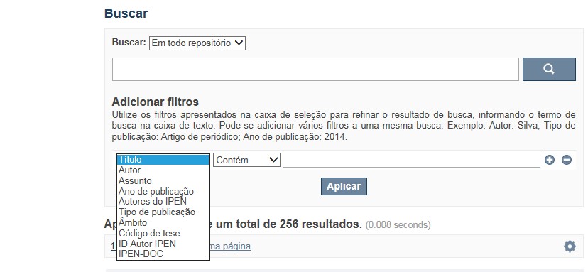
A opção Busca avançada utiliza os conectores da lógica boleana, é o melhor recurso para combinar chaves de busca e obter documentos relevantes à sua pesquisa, utilize os filtros apresentados na caixa de seleção para refinar o resultado de busca. Pode-se adicionar vários filtros a uma mesma busca.
Exemplo:
Buscar os artigos apresentados em um evento internacional de 2015, sobre loss of coolant, do autor Maprelian.
Autor: Maprelian
Título: loss of coolant
Tipo de publicação: Texto completo de evento
Ano de publicação: 2015

✔ Para indexação dos documentos é utilizado o Thesaurus do INIS, especializado na área nuclear e utilizado em todos os países membros da International Atomic Energy Agency – IAEA , por esse motivo, utilize os termos de busca de assunto em inglês; isto não exclui a busca livre por palavras, apenas o resultado pode não ser tão relevante ou pertinente.
✔ 95% do RD apresenta o texto completo do documento com livre acesso, para aqueles que apresentam o ![]() significa que e o documento está sujeito as leis de direitos autorais, solicita-se nesses casos contatar a Biblioteca do IPEN,
bibl@ipen.br
.
significa que e o documento está sujeito as leis de direitos autorais, solicita-se nesses casos contatar a Biblioteca do IPEN,
bibl@ipen.br
.
✔ Ao efetuar a busca por um autor o RD apresentará uma relação de todos os trabalhos depositados no RD. No lado direito da tela são apresentados os coautores com o número de trabalhos produzidos em conjunto bem como os assuntos abordados e os respectivos anos de publicação agrupados.
✔ O RD disponibiliza um quadro estatístico de produtividade, onde é possível visualizar o número dos trabalhos agrupados por tipo de coleção, a medida que estão sendo depositados no RD.
✔ Na página inicial nas referências são sinalizados todos os autores IPEN, ao clicar nesse símbolo ![]() será aberta uma nova página correspondente à aquele autor – trata-se da página do pesquisador.
será aberta uma nova página correspondente à aquele autor – trata-se da página do pesquisador.
✔ Na página do pesquisador, é possível verificar, as variações do nome, a relação de todos os trabalhos com texto completo bem como um quadro resumo numérico; há links para o Currículo Lattes e o Google Acadêmico ( quando esse for informado).
ATENÇÃO!
ESTE TEXTO "AJUDA" ESTÁ SUJEITO A ATUALIZAÇÕES CONSTANTES, A MEDIDA QUE NOVAS FUNCIONALIDADES E RECURSOS DE BUSCA FOREM SENDO DESENVOLVIDOS PELAS EQUIPES DA BIBLIOTECA E DA INFORMÁTICA.
O gerenciamento do Repositório está a cargo da Biblioteca do IPEN. Constam neste RI, até o presente momento 20.950 itens que tanto podem ser artigos de periódicos ou de eventos nacionais e internacionais, dissertações e teses, livros, capítulo de livros e relatórios técnicos. Para participar do RI-IPEN é necessário que pelo menos um dos autores tenha vínculo acadêmico ou funcional com o Instituto. Nesta primeira etapa de funcionamento do RI, a coleta das publicações é realizada periodicamente pela equipe da Biblioteca do IPEN, extraindo os dados das bases internacionais tais como a Web of Science, Scopus, INIS, SciElo além de verificar o Currículo Lattes. O RI-IPEN apresenta também um aspecto inovador no seu funcionamento. Por meio de metadados específicos ele está vinculado ao sistema de gerenciamento das atividades do Plano Diretor anual do IPEN (SIGEPI). Com o objetivo de fornecer dados numéricos para a elaboração dos indicadores da Produção Cientifica Institucional, disponibiliza uma tabela estatística registrando em tempo real a inserção de novos itens. Foi criado um metadado que contém um número único para cada integrante da comunidade científica do IPEN. Esse metadado se transformou em um filtro que ao ser acionado apresenta todos os trabalhos de um determinado autor independente das variáveis na forma de citação do seu nome.
A elaboração do projeto do RI do IPEN foi iniciado em novembro de 2013, colocado em operação interna em julho de 2014 e disponibilizado na Internet em junho de 2015. Utiliza o software livre Dspace, desenvolvido pelo Massachusetts Institute of Technology (MIT). Para descrição dos metadados adota o padrão Dublin Core. É compatível com o Protocolo de Arquivos Abertos (OAI) permitindo interoperabilidade com repositórios de âmbito nacional e internacional.
1. Portaria IPEN-CNEN/SP nº 387, que estabeleceu os princípios que nortearam a criação do RDI, clique aqui.
2. A experiência do Instituto de Pesquisas Energéticas e Nucleares (IPEN-CNEN/SP) na criação de um Repositório Digital Institucional – RDI, clique aqui.
O Repositório Digital do IPEN é um equipamento institucional de acesso aberto, criado com o objetivo de reunir, preservar, disponibilizar e conferir maior visibilidade à Produção Científica publicada pelo Instituto, desde sua criação em 1956.
Operando, inicialmente como uma base de dados referencial o Repositório foi disponibilizado na atual plataforma, em junho de 2015. No Repositório está disponível o acesso ao conteúdo digital de artigos de periódicos, eventos, nacionais e internacionais, livros, capítulos, dissertações, teses e relatórios técnicos.
A elaboração do projeto do RI do IPEN foi iniciado em novembro de 2013, colocado em operação interna em julho de 2014 e disponibilizado na Internet em junho de 2015. Utiliza o software livre Dspace, desenvolvido pelo Massachusetts Institute of Technology (MIT). Para descrição dos metadados adota o padrão Dublin Core. É compatível com o Protocolo de Arquivos Abertos (OAI) permitindo interoperabilidade com repositórios de âmbito nacional e internacional.
O gerenciamento do Repositório está a cargo da Biblioteca do IPEN. Constam neste RI, até o presente momento 20.950 itens que tanto podem ser artigos de periódicos ou de eventos nacionais e internacionais, dissertações e teses, livros, capítulo de livros e relatórios técnicos. Para participar do RI-IPEN é necessário que pelo menos um dos autores tenha vínculo acadêmico ou funcional com o Instituto. Nesta primeira etapa de funcionamento do RI, a coleta das publicações é realizada periodicamente pela equipe da Biblioteca do IPEN, extraindo os dados das bases internacionais tais como a Web of Science, Scopus, INIS, SciElo além de verificar o Currículo Lattes. O RI-IPEN apresenta também um aspecto inovador no seu funcionamento. Por meio de metadados específicos ele está vinculado ao sistema de gerenciamento das atividades do Plano Diretor anual do IPEN (SIGEPI). Com o objetivo de fornecer dados numéricos para a elaboração dos indicadores da Produção Cientifica Institucional, disponibiliza uma tabela estatística registrando em tempo real a inserção de novos itens. Foi criado um metadado que contém um número único para cada integrante da comunidade científica do IPEN. Esse metadado se transformou em um filtro que ao ser acionado apresenta todos os trabalhos de um determinado autor independente das variáveis na forma de citação do seu nome.
