Navegação Periódicos - Resumos por assunto "lasers"
- Página inicial
- →
- IPEN
- →
- Periódicos - Resumos
- →
- Navegação Periódicos - Resumos por assunto
- Sobre
- Perfil Técnico
- Política de funcionamento
- Ajuda
- Apresentação
Navegação Periódicos - Resumos por assunto "lasers"
Itens para a visualização no momento 1-20 de 23
-
. Bacterial reduction by photodynamic therapy in peri-implantitis: an in vivo study. Brazilian Dental Journal, v. 15, Special issue, p. 84-84, 2004. Abstract: Progressive peri-implantar bone losses, which are accompanied by inflammatory process in the soft tissues is referred to as peri-implantitis. The aim of this study was to compare the effects of lethal photosensitization with the conventional technique on bacterial reduction in ligature induced peri-implantitis in dogs. Seventeen third premolars of eight Labrador dogs were extracted and, immediately after, the implants were submerged. After osseointegration, peri-implantitis was induced. After 4 months, ligature were removed and the same period was waited for natural induction of bacterial plaque. The dogs were randomly divided into two groups. In the conventional group, they were treated with the conventional techniques of mucoperiosteal flaps for scaling the implant surface and irrigation with chlorexidine. In the laser group, only mucoperiosteal scaling was carried out before photodynamic therapy. On the peri-implantar pocket an azulene paste was introduced and a GaAlAs low-power laser (l= 660 nm, P= 30 mW, E= 5,4 J and Dt= 3 min.) was applied. Microbiological samples were obtained before and immediately after treatment. The results of this study showed that Prevotella sp., Fusobacterium e S. Beta-haemolyticus were significantly reduced for the conventional and laser groups (100%,99.8%; 100%,100%; 85.7%,97.6%, respectively).
Palavras-Chave: dentistry; teeth; implants; bone tissues; bacteria; photodynamic therapy; lasers; photosensitivity
. Bacterial reduction by photodynamic therapy in peri-implantitis: an in vivo study. Brazilian Dental Journal, v. 15, p. 84-84, 2004. Special issue. Disponível em: http://repositorio.ipen.br/handle/123456789/32755. Acesso em: $DATA.Como referenciar este itemEsta referência é gerada automaticamente de acordo com as normas do estilo IPEN/SP (ABNT NBR 6023) e recomenda-se uma verificação final e ajustes caso necessário.
-
. Bacterial reduction in class II furcation after root debridment with or without Nd:YAG laser irradiation. Brazilian Dental Journal, v. 15, Special issue, p. 90-91, 2004. Abstract: The use of Nd:Yag laser for bacterial reduction as an adjuvant to nonsurgical periodontal treatment has been approached in several studies. Furcation complex anatomy is responsible for comprised treatment results in this areas due to the lack of proper access for instrumentation showing the persistence of a pathogenic microbial flora. The purpose of this clinical trial, randomized, double-blinded was to evaluated the bacterial reduction achieved with the Nd:YAG associated to conventional treatment on furcation sites of patients with chronic periodontitis. In a split mouth design study, 34 class II furcations that were selected from 17 patients with chronic periodontitis. They received previous full mouth periodontal treatment, except for the experimental sites. The 17 furcations of the Control group underwent twice manual and ultrasonic root debridment in weekly intervals. The Test group received the same treatment as the Control group followed by the Nd:YAG laser application (100mJ/pulse; 1.5W; 15Hz; 60sec). The microbiological parameters total numbers of anaerobic Colony Forming Units(CFU); Black pigmented CFU and the level of Actinobacillus actinomycetemcomitans(Aa), Porphyromonas gingivalis (Pg) and Prevotella intermedia(Pi) were determined at baseline, immediatly and one month after the treatment. The results showed a significant reduction of total CFU for both groups immediately after the treatment, but it was better for the Test group. After one month the total CFU average increased but was still below pretreatment levels for both groups. The black pigmented CFU and the level of Aa, Pg e Pi decreased significantly after the treatment but 30 days after there was an increase almost equal to baseline levels for both groups. The Nd:Yag laser associated with convencional treatment promoted bacterial reduction on class II furcation immediately after its application.
Palavras-Chave: bacteria; laser radiation; lasers; clinical trials
. Bacterial reduction in class II furcation after root debridment with or without Nd:YAG laser irradiation. Brazilian Dental Journal, v. 15, p. 90-91, 2004. Special issue. Disponível em: http://repositorio.ipen.br/handle/123456789/32740. Acesso em: $DATA.Como referenciar este itemEsta referência é gerada automaticamente de acordo com as normas do estilo IPEN/SP (ABNT NBR 6023) e recomenda-se uma verificação final e ajustes caso necessário.
-
. Carbon dioxide laser or cold scalpel on the removal of gingival melanin pigmentation: comparative study. Brazilian Dental Journal, v. 15, Special issue, p. 90-90, 2004. Abstract: Melanin pigmentation is the result of melanin granules produced by melanocytes present in the basal layer of the oral epithelium. Gingival physiological melanin pigmentation is symmetric and persistent, may cause esthetic problems especially in individuals with a gummy smile. Various techniques have been described for the removal of melanin pigmentation from the gingival epithelium and partial thin connective tissue, as chemical agents, cryosurgery, surgery and gingival grafts. Recently, lasers systems have been used to coagulate and vaporize cells, promoting controlled gingival ablation. This study compares clinical efficiency to removal gingival melanin pigmentation in 20 patients with dioxide carbon laser, and 20 patients with cold scalpel during 30 days after surgery. A dioxide carbon laser (output = 5W; superpulse = 0,5s; spot size = 2,5mm defocused; focal distance = 5,5cm, Intensity = 102 W/cm2) was irradiated on gingival mucosal surface. Both techniques presented epithelialization in 15 days. Both systems are considered effective for removal melanin pigments. Patient's evaluation with postoperative pain found the carbon dioxide laser technique superior to the cold scalpel one. After 30 days, the repigmentation occured in 45% of the dioxide carbon laser patients, and 80% of the cold scalpel patients.
Palavras-Chave: carbon dioxide lasers; melanin; pigments; lasers
. Carbon dioxide laser or cold scalpel on the removal of gingival melanin pigmentation: comparative study. Brazilian Dental Journal, v. 15, p. 90-90, 2004. Special issue. Disponível em: http://repositorio.ipen.br/handle/123456789/32739. Acesso em: $DATA.Como referenciar este itemEsta referência é gerada automaticamente de acordo com as normas do estilo IPEN/SP (ABNT NBR 6023) e recomenda-se uma verificação final e ajustes caso necessário.
-
. Caries-preventive effect of infrared lasers and professional fluoride application on enamel. Caries Research, v. 43, p. 192, 2009.
Palavras-Chave: caries; preventive medicine; infrared radiation; lasers; calcium fluorides; enamels
. Caries-preventive effect of infrared lasers and professional fluoride application on enamel. Caries Research, v. 43, p. 192, 2009. Disponível em: http://repositorio.ipen.br/handle/123456789/8826. Acesso em: $DATA.Como referenciar este itemEsta referência é gerada automaticamente de acordo com as normas do estilo IPEN/SP (ABNT NBR 6023) e recomenda-se uma verificação final e ajustes caso necessário.
-
. Cavity preparation with ER:YAG laser: pain evaluation. Brazilian Dental Journal, v. 15, Special issue, p. 94-94, 2004. Abstract: They were selected for this work clinic patient of the which were selected 15 teeth with decay lesion, being ten teeth with lesion type class I, of these five for the group-control with high conventional rotation, and five for the group laser class I, and five teeth with lesion type class Vfor the group laser. In the preparations with laser of Er:YAG (Kavo Key Laser 2), any patient do not was anesthetized, even in the deepest cavities, and the maximum degree of pain (that varied from 0 to 10) it was of 4. In the group-control, with mounted tip in high conventional rotation, two patients were anesthetized, and the maximum degree of pain was of 7. The use of the laser in the dental clinic (restorative dentistry), using the technology laser in the dental preparations, it showed to be a good alternative to the use of the mounted tip in high conventional rotation. 94
Palavras-Chave: dentin; teeth; oral cavity; lasers; biological recovery; pain
. Cavity preparation with ER:YAG laser: pain evaluation. Brazilian Dental Journal, v. 15, p. 94-94, 2004. Special issue. Disponível em: http://repositorio.ipen.br/handle/123456789/32743. Acesso em: $DATA.Como referenciar este itemEsta referência é gerada automaticamente de acordo com as normas do estilo IPEN/SP (ABNT NBR 6023) e recomenda-se uma verificação final e ajustes caso necessário.
-
. Changes in chemical composition and collagen structure of dentin tissue after erbium laser irradiation. Brazilian Dental Journal, v. 15, Special issue, p. 78-78, 2004. Abstract: The erbium laser light has a great affinity to the water molecule, which is present in great quantity in biological hard tissues. The objective of this work is to identify chemical changes by infrared spectroscopy of irradiated dentin by an Er:YAG - 2.94μm laser. The irradiation was performed with fluences between 0.365 J/cm2 and 1.94 J/cm2. For the infrared analysis a Fourier transform infrared spectrometer was used. After the irradiation were observed: loss of water, alteration of the structure and composition of the collagen and increase of the OH- radical. These alterations can be identified by a decrease of the water and OH- band between 3800-2800 cm-1, bands ascribed to collagen structure between 1400-1100 cm-1. The results show that the erbium laser changes the structure and composition of the organic matrix, OHradical and the water composition in the irradiated dentin.
Palavras-Chave: dentin; bone tissues; teeth; erbium; lasers
. Changes in chemical composition and collagen structure of dentin tissue after erbium laser irradiation. Brazilian Dental Journal, v. 15, p. 78-78, 2004. Special issue. Disponível em: http://repositorio.ipen.br/handle/123456789/32734. Acesso em: $DATA.Como referenciar este itemEsta referência é gerada automaticamente de acordo com as normas do estilo IPEN/SP (ABNT NBR 6023) e recomenda-se uma verificação final e ajustes caso necessário.
-
. Comparative study of action by etching modalities and Er:YAG laser irradiation on the root surface. Journal of Dental Research, v. 81, p. 195, 2002.
Palavras-Chave: lasers; erbium; irradiation; citric acid; edta; comparative evaluations; surfaces; oral cavity; in vitro; scanning electron microscopy
. Comparative study of action by etching modalities and Er:YAG laser irradiation on the root surface. Journal of Dental Research, v. 81, p. 195, 2002. Disponível em: http://repositorio.ipen.br/handle/123456789/8787. Acesso em: $DATA.Como referenciar este itemEsta referência é gerada automaticamente de acordo com as normas do estilo IPEN/SP (ABNT NBR 6023) e recomenda-se uma verificação final e ajustes caso necessário.
-
. Effect of Er:YAG and diode lasers in the adhesion of blood components and in the morphology of irradiated root surfaces. Brazilian Dental Journal, v. 15, Special issue, p. 89-89, 2004. Abstract: The aim of this study was to evaluate in vitro the adhesion of blood components on root surfaces irradiated with Er:YAG (2.94??m) and GaAlAs Diode (808 nm) lasers and these effects on irradiated root surfaces. It was obtained 100 samples of human teeth. They were scaled and divided into five groups of 20 samples each: G1 (Control); G2 -Er:YAG laser (7.6 J/cm2); G3 - Er:YAG laser (12.9 J/cm2); G4 -Diode laser (90 J/cm2) and G5 - Diode laser (108 J/cm2). After these treatments were conducted, 10 samples of each group received a blood tissue, and the reminiscent 10 samples did not receive such treatment. After laboratorial treatments the samples were analysed by scanning electron microscopy. The results have shown that there were no significant differences between the Control Group and the groups treated with Er:YAG laser (p=0,9633 and 0,6229); G4 and G5 were less effective than the Control Group and the Er:YAG laser groups (p<0,01). No proposed treatment increased the adhesion of blood components in a significant way when compared to the Control Group; although the Er:YAG laser did not interfere in the adhesion of blood components it caused more changes on the root surface, while the Diode laser inhibited the adhesion.
Palavras-Chave: teeth; blood; fibrin; lasers; erbium; in vitro; morphological changes
. Effect of Er:YAG and diode lasers in the adhesion of blood components and in the morphology of irradiated root surfaces. Brazilian Dental Journal, v. 15, p. 89-89, 2004. Special issue. Disponível em: http://repositorio.ipen.br/handle/123456789/32737. Acesso em: $DATA.Como referenciar este itemEsta referência é gerada automaticamente de acordo com as normas do estilo IPEN/SP (ABNT NBR 6023) e recomenda-se uma verificação final e ajustes caso necessário.
-
. Effect of infrared lasers on chemical and crystalline properties of enamel. Caries Research, v. 43, p. 192, 2009.
Palavras-Chave: caries; preventive medicine; infrared radiation; lasers; enamels; radiation effects; chemical properties; morphological changes
. Effect of infrared lasers on chemical and crystalline properties of enamel. Caries Research, v. 43, p. 192, 2009. Disponível em: http://repositorio.ipen.br/handle/123456789/8827. Acesso em: $DATA.Como referenciar este itemEsta referência é gerada automaticamente de acordo com as normas do estilo IPEN/SP (ABNT NBR 6023) e recomenda-se uma verificação final e ajustes caso necessário.
-
. Effects of He-Ne Laser on blood microcirculation during wound healing process. Lasers in Surgery and Medicine, v. suppl. 16, p. 4, 2004.
Palavras-Chave: blood flow; flowmeters; therapy; skin; repair; lasers; doppler effect
. Effects of He-Ne Laser on blood microcirculation during wound healing process. Lasers in Surgery and Medicine, v. suppl. 16, p. 4, 2004. Disponível em: http://repositorio.ipen.br/handle/123456789/8809. Acesso em: $DATA.Como referenciar este itemEsta referência é gerada automaticamente de acordo com as normas do estilo IPEN/SP (ABNT NBR 6023) e recomenda-se uma verificação final e ajustes caso necessário.
-
. Effects of low intensity laser radiation in the prevention of oral mucositis in patients undergoing bone marrow transplant. Brazilian Dental Journal, v. 15, Special issue, p. 97-97, 2004. Abstract: Oral mucositis is one of the complications arising from pre bone marrow transplant conditioning, which can substantially change the patient's quality of life. The purpose of this randomized double blind study was to compare the effects of low intensity laser radiation in the prevention of oral mucositis in patients undergoing bone marrow transplants. Seventy patients at the Seattle Cancer Care Alliance in the U.S.A. were approved by the local ethics committee and gave their informed consent to take part in the study. The 70 patients were divided into three groups (group 1 - laser 650nm; group 2 - laser 780nm and group 3 placebo). The therapy or placebo treatment began on the first day of the conditioning and continued through to two days following the bone marrow transplant. Mucositis was measured according to the oral mucositis rate and the pain assessment rate (VAS). We were thus able to conclude that the diode 650nm laser indeed decreased the severity of oral mucositis as well as the degree the pain when used as a preventative therapy in patients undergoing bone marrow transplants. In this study, low intensity laser therapy was regarded as safe and did not present any side effects.
Palavras-Chave: low dose irradiation; oral cavity; mucous membranes; lasers; semiconductor diodes; transplants; bone marrow
. Effects of low intensity laser radiation in the prevention of oral mucositis in patients undergoing bone marrow transplant. Brazilian Dental Journal, v. 15, p. 97-97, 2004. Special issue. Disponível em: http://repositorio.ipen.br/handle/123456789/32744. Acesso em: $DATA.Como referenciar este itemEsta referência é gerada automaticamente de acordo com as normas do estilo IPEN/SP (ABNT NBR 6023) e recomenda-se uma verificação final e ajustes caso necessário.
-
. Effects of low-intensity red laser radiation on the dentine-pulp interface after class I cavity preparation disfunction. Brazilian Dental Journal, v. 15, Special issue, p. 86-87, 2004. Abstract: The aim of this study was to investigate the effects of low-intensity red laser radiation on the ultrastructure of dentine-pulp interface after conventionally prepared class I cavity preparation. Eight premolars indicated for extraction for orthodontic reasons from 2 patients were used. Class I cavities were prepared and the teeth were divided into two groups. The first group received a treatment with a GaAlAs laser, l= 660 nm, P= 30 mW and D= 2J/cm2. The laser tip was applied directly and perpendicularly into the cavity in only one sense. The teeth from the second group had their class I cavities prepared but they did not receive the laser therapy. All cavities were filled with composite resin. Twenty-eight days after the preparation, the teeth were extracted and processed for transmission electron microscopy analysis. Two sound teeth (healthy group) without any preparation were also examined. The first group presented odontoblastic processes in intimate contact with the extracellular matrix, while the collagen fibers appeared more aggregated and organized than those of the second group. These results were also observed in the healthy teeth. The results suggest that laser irradiation accelerates the recovery of the structures at the dentine-pulp interface involved during cavity preparation layer.
Palavras-Chave: teeth; dentin; oral cavity; lasers; radiation effects
. Effects of low-intensity red laser radiation on the dentine-pulp interface after class I cavity preparation disfunction. Brazilian Dental Journal, v. 15, p. 86-87, 2004. Special issue. Disponível em: http://repositorio.ipen.br/handle/123456789/32758. Acesso em: $DATA.Como referenciar este itemEsta referência é gerada automaticamente de acordo com as normas do estilo IPEN/SP (ABNT NBR 6023) e recomenda-se uma verificação final e ajustes caso necessário.
-
. Er,Cr:YSGG laser irradiation associated to fluoride for in situ model using gamma sterilized dentin and enamel. Photobiomodulation, Photomedicine, and Laser Surgery, v. 37, n. 10, p. A13-A13, 2019. DOI: 10.1089/photob.2019.29013.abstracts Abstract: The in situ intraoral model uses human dental enamel samples (HDE) in order to analyse the de-remineralization processes using the buccal environment without interfering into the patients’ natural dentition. The main ethical concern from this model is the biosafety. Gamma radiation is a very efficient sterilization method that is not expected to alter the mineral content of the hard tissues, avoiding biases in the results. Thus 40 HDE samples were irradiated through a source of 60Co multipurpose irradiator aiming complete sterilization (25 KGy/h) with the purpose of accumulating the native plaque on them at an in situ study. An Er,Cr:YSGG laser was used alone and in combination with the topical applications of: 1-dentifrice (1,100 lg F-/g) or 2-APF (12,300 lg F-/g). Morphological analyses were performed by scanning electron microscopy (SEM), determination of alkali-soluble fluoride concentration by specific ion electrode and microhardness determination. Then, the 15 volunteers used palatal devices containing previously treated HDE samples and remained using F dentifrice. The FTIR findings established that gamma radiation could be used aiming HDE sterilization. The Knoop hardness number was within the range of that of natural dentin of human origin. X-ray fluorescence shows that irradiated dentin has great similarity with natural dentin from the point of view of chemical composition. SEM analyses showed that there was no thermal damage or interprismatic morphological changes in the hydroxyapatite structure of human dental dentin outside the buccal environment when using doses of gamma irradiation up to 25 kGy.
Palavras-Chave: teeth; enamels; sterilization; lasers; irradiation
. Er,Cr:YSGG laser irradiation associated to fluoride for in situ model using gamma sterilized dentin and enamel. Photobiomodulation, Photomedicine, and Laser Surgery, v. 37, n. 10, p. A13-A13, 2019. DOI: 10.1089/photob.2019.29013.abstracts. Disponível em: http://repositorio.ipen.br/handle/123456789/32221. Acesso em: $DATA.Como referenciar este itemEsta referência é gerada automaticamente de acordo com as normas do estilo IPEN/SP (ABNT NBR 6023) e recomenda-se uma verificação final e ajustes caso necessário.
-
. Esmalte sadio e cariado irradiado com laserterapia infravermelho e creme: Estudo da microdureza, temperatura superficial e pulpar. Brazilian Oral Research, v. 25, n. Supl. 1, p. 297, 2011.
Palavras-Chave: teeth; caries; enamels; lasers; therapy; abrasives; fluorides; infrared spectra; microhardness; temperature dependence
. Esmalte sadio e cariado irradiado com laserterapia infravermelho e creme: Estudo da microdureza, temperatura superficial e pulpar. Brazilian Oral Research, v. 25, n. Supl. 1, p. 297, 2011. Disponível em: http://repositorio.ipen.br/handle/123456789/8888. Acesso em: $DATA.Como referenciar este itemEsta referência é gerada automaticamente de acordo com as normas do estilo IPEN/SP (ABNT NBR 6023) e recomenda-se uma verificação final e ajustes caso necessário.
-
. Fluoride incorporation and acid resistance of dental enamel irradiated with Er:YAG: atomic absorption spectrometry and spectrophotometry. Brazilian Dental Journal, v. 15, Special issue, p. 131-131, 2004. Abstract: Er:YAG effects on dental enamel surface regarding the resistance to demineralization and the fluoride incorporation were evaluated. 80 samples were divided into 8 groups: G1) control - APF application; G2) conditioning with 37% phosphoric acid and APF application; G3) irradiation with 250 mJ/pulse, 7 Hz, 31,84 J/cm2 (contact) and APF application; G4) irradiation with 200 mJ/pulse, 7 Hz, 25,47 J/cm2 (contact) and APF application; G5) irradiation with 150 mJ/pulse, 7 Hz, 19,10 J/cm2 (contact) and APF application; G6) irradiation with 250 mJ/pulse, 7 Hz, 2,08 J/cm2 (non-contact) and APF application; G7) irradiation with 200 mJ/pulse, 7 Hz, 1,8 J/cm2 (non-contact) and APF application; G8) irradiation with 100 mJ/pulse, 7 Hz, 0,9 J/cm2 (noncontact) and APF application. All samples were immersed in 2,0 M acetic-acetate acid solution, pH 4,5 for 8 hours. The fluoride, calcium and phosphorous ions were analyzed, by atomic absorption spectrometry and spectrophotometry. Groups laser irradiated before topic APF application presented better results than the control. There was higher fluoride incorporation on G7 and G8. Calcium and phosphorous analysis reveled a decrease on the enamel demineralization on G2 and G3 groups. The Er:YAG laser on irradiation conditions of this work is a promissory alternative for the Preventive Dentistry.
Palavras-Chave: dentistry; enamels; lasers; laser radiation; fluorides
. Fluoride incorporation and acid resistance of dental enamel irradiated with Er:YAG: atomic absorption spectrometry and spectrophotometry. Brazilian Dental Journal, v. 15, p. 131-131, 2004. Special issue. Disponível em: http://repositorio.ipen.br/handle/123456789/32751. Acesso em: $DATA.Como referenciar este itemEsta referência é gerada automaticamente de acordo com as normas do estilo IPEN/SP (ABNT NBR 6023) e recomenda-se uma verificação final e ajustes caso necessário.
-
. Focus issue introduction: Advanced Solid-State Lasers 2020. Optics Express, v. 29, n. 6, p. 8365-8367, 2021. DOI: 10.1364/OE.423636 Abstract: This Joint Issue of Optics Express and Optical Materials Express features 15 articles written by authors who participated in the international online conference Advanced Solid State Lasers held 13–16 October, 2020. This review provides a summary of the conference and these articles from the conference which sample the spectrum of solid state laser theory and experiment, from materials research to sources and from design innovation to applications.
Palavras-Chave: lasers; solid state lasers; radiation sources; meetings
. Focus issue introduction: Advanced Solid-State Lasers 2020. Optics Express, v. 29, n. 6, p. 8365-8367, 2021. DOI: 10.1364/OE.423636. Disponível em: http://repositorio.ipen.br/handle/123456789/32049. Acesso em: $DATA.Como referenciar este itemEsta referência é gerada automaticamente de acordo com as normas do estilo IPEN/SP (ABNT NBR 6023) e recomenda-se uma verificação final e ajustes caso necessário.
-
. Focus issue introduction: Advanced Solid-State Lasers 2020. Optical Materials Express, v. 11, n. 4, p. 952-954, 2021. DOI: 10.1364/OME.423641 Abstract: This Joint Issue of Optics Express and Optical Materials Express features 15 articles written by authors who participated in the international online conference Advanced Solid State Lasers held 13–16 October, 2020. This review provides a summary of the conference and these articles from the conference which sample the spectrum of solid state laser theory and experiment, from materials research to sources and from design innovation to applications.
Palavras-Chave: lasers; solid state lasers; radiation sources; meetings
. Focus issue introduction: Advanced Solid-State Lasers 2020. Optical Materials Express, v. 11, n. 4, p. 952-954, 2021. DOI: 10.1364/OME.423641. Disponível em: http://repositorio.ipen.br/handle/123456789/32048. Acesso em: $DATA.Como referenciar este itemEsta referência é gerada automaticamente de acordo com as normas do estilo IPEN/SP (ABNT NBR 6023) e recomenda-se uma verificação final e ajustes caso necessário.
-
. In vitro thermographic measurement in pulpal chamber during diode laser bleaching. Brazilian Dental Journal, v. 15, Special issue, p. 94-94, 2004. Abstract: Thermographic was employed to determine the temperature rise in lower incisors pulpal chambers during diode laser bleaching. Two methods were used: a thermocouple for 72 teeth and a infrared (IR) thermographic camera for 36. Two bleaching agents, both 35% hydrogen peroxide- Whiteness HP (HP) and Hi Lite (HL) - were applied to the specimens buccal surfaces and irradiated with a diode laser (808 5nm), CW for 30s. Intensities tested were 21.W/cm2, 29.8W/cm2, 35.8W/cm2, 38.2W/cm2, 52.9W/cm2 e 63.7W/cm2. Means of the greatest temperature rises with the HL were statiscally lower than the HP (p<0.01). When HP was irradiated with 50.9W/cm² and 61.1W/cm², the temperature registered was over 5.5ºC, considered as the limit to avoid pulp damage. The IR thermacam analysis showed that, when the HP was used, the temperature rise in pulp chamber was similar to the target area on the buccal surface. Evaluation of tooth color was done using a VITAshade guide at baseline and at the end of the bleaching treatment. Both products proved to be efficient, however HP produced statiscally higher shade changes than HL (p<0.01). It can be concluded that the diode laser bleaching associated with the HP was safe when intensities below 50mW/cm2 were employed. Higher parameters can cause damage to pulp vitality of the lower incisors, fact that did not occurred with the HL gel. Both gels were efficacious to the bleaching technique proposed, but the HP showed better results.
Palavras-Chave: dentin; teeth; bleaching; thermography; hydrogen peroxide; semiconductor diodes; lasers
. In vitro thermographic measurement in pulpal chamber during diode laser bleaching. Brazilian Dental Journal, v. 15, p. 94-94, 2004. Special issue. Disponível em: http://repositorio.ipen.br/handle/123456789/32742. Acesso em: $DATA.Como referenciar este itemEsta referência é gerada automaticamente de acordo com as normas do estilo IPEN/SP (ABNT NBR 6023) e recomenda-se uma verificação final e ajustes caso necessário.
-
. Laser panorama in dentistry in the international year of light / Panorama do laser em odontologia no ano internacional da luz. Brazilian Dental Science, v. 18, n. 4, p. 3, 2015.
Palavras-Chave: lasers; dentistry; uses; therapy
. Laser panorama in dentistry in the international year of light. Brazilian Dental Science, v. 18, n. 4, p. 3, 2015. Disponível em: http://repositorio.ipen.br/handle/123456789/26551. Acesso em: $DATA.Como referenciar este itemEsta referência é gerada automaticamente de acordo com as normas do estilo IPEN/SP (ABNT NBR 6023) e recomenda-se uma verificação final e ajustes caso necessário.
-
. Latin America Optics and Photonics 2022: introduction to the feature issue. Applied Optics, v. 62, n. 8, p. 1-2, 2023. DOI: 10.1364/AO.489414 Abstract: The 2022 Latin America Optics and Photonics Conference (LAOP 2022), the major international conference sponsored byOptica in Latin America, returned to Recife, Pernambuco, Brazil, after its first edition in 2010.Held every two years since (except for 2020), LAOP has the explicit objective to promote Latin American excellence in optics and photonics research and support the regional community. In the 6th edition in 2022, it featured a comprehensive technical program with recognized experts in fields critical to Latin America, highly multidisciplinary, with themes frombiophotonics to2Dmaterials. The 191 attendees of LAOP2022 listened to five plenary speakers, 28 keynotes, 24 invited talks, and 128 presentations, including oral and posters
Palavras-Chave: optical activity; photon emission; lasers; fiber optics; meetings
. Latin America Optics and Photonics 2022: introduction to the feature issue. Applied Optics, v. 62, n. 8, p. 1-2, 2023. DOI: 10.1364/AO.489414. Disponível em: http://repositorio.ipen.br/handle/123456789/34097. Acesso em: $DATA.Como referenciar este itemEsta referência é gerada automaticamente de acordo com as normas do estilo IPEN/SP (ABNT NBR 6023) e recomenda-se uma verificação final e ajustes caso necessário.
Itens para a visualização no momento 1-20 de 23
Buscar no repositório
Navegar
Minha conta
Visualizar
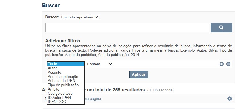
A pesquisa no RD utiliza os recursos de busca da maioria das bases de dados. No entanto algumas dicas podem auxiliar para obter um resultado mais pertinente.
✔ É possível efetuar a busca de um autor ou um termo em todo o RD, por meio do Buscar no Repositório , isto é, o termo solicitado será localizado em qualquer campo do RD. No entanto esse tipo de pesquisa não é recomendada a não ser que se deseje um resultado amplo e generalizado.
✔ A pesquisa apresentará melhor resultado selecionando um dos filtros disponíveis em Navegar
✔ Os filtros disponíveis em Navegar tais como: Coleções, Ano de publicação, Títulos, Assuntos, Autores, Revista, Tipo de publicação são autoexplicativos. O filtro, Autores IPEN apresenta uma relação com os autores vinculados ao IPEN; o ID Autor IPEN diz respeito ao número único de identificação de cada autor constante no RD e sob o qual estão agrupados todos os seus trabalhos independente das variáveis do seu nome; Tipo de acesso diz respeito à acessibilidade do documento, isto é , sujeito as leis de direitos autorais, ID RT apresenta a relação dos relatórios técnicos, restritos para consulta das comunidades indicadas.

A opção Busca avançada utiliza os conectores da lógica boleana, é o melhor recurso para combinar chaves de busca e obter documentos relevantes à sua pesquisa, utilize os filtros apresentados na caixa de seleção para refinar o resultado de busca. Pode-se adicionar vários filtros a uma mesma busca.
Exemplo:
Buscar os artigos apresentados em um evento internacional de 2015, sobre loss of coolant, do autor Maprelian.
Autor: Maprelian
Título: loss of coolant
Tipo de publicação: Texto completo de evento
Ano de publicação: 2015

✔ Para indexação dos documentos é utilizado o Thesaurus do INIS, especializado na área nuclear e utilizado em todos os países membros da International Atomic Energy Agency – IAEA , por esse motivo, utilize os termos de busca de assunto em inglês; isto não exclui a busca livre por palavras, apenas o resultado pode não ser tão relevante ou pertinente.
✔ 95% do RD apresenta o texto completo do documento com livre acesso, para aqueles que apresentam o ![]() significa que e o documento está sujeito as leis de direitos autorais, solicita-se nesses casos contatar a Biblioteca do IPEN,
bibl@ipen.br
.
significa que e o documento está sujeito as leis de direitos autorais, solicita-se nesses casos contatar a Biblioteca do IPEN,
bibl@ipen.br
.
✔ Ao efetuar a busca por um autor o RD apresentará uma relação de todos os trabalhos depositados no RD. No lado direito da tela são apresentados os coautores com o número de trabalhos produzidos em conjunto bem como os assuntos abordados e os respectivos anos de publicação agrupados.
✔ O RD disponibiliza um quadro estatístico de produtividade, onde é possível visualizar o número dos trabalhos agrupados por tipo de coleção, a medida que estão sendo depositados no RD.
✔ Na página inicial nas referências são sinalizados todos os autores IPEN, ao clicar nesse símbolo ![]() será aberta uma nova página correspondente à aquele autor – trata-se da página do pesquisador.
será aberta uma nova página correspondente à aquele autor – trata-se da página do pesquisador.
✔ Na página do pesquisador, é possível verificar, as variações do nome, a relação de todos os trabalhos com texto completo bem como um quadro resumo numérico; há links para o Currículo Lattes e o Google Acadêmico ( quando esse for informado).
ATENÇÃO!
ESTE TEXTO "AJUDA" ESTÁ SUJEITO A ATUALIZAÇÕES CONSTANTES, A MEDIDA QUE NOVAS FUNCIONALIDADES E RECURSOS DE BUSCA FOREM SENDO DESENVOLVIDOS PELAS EQUIPES DA BIBLIOTECA E DA INFORMÁTICA.
O gerenciamento do Repositório está a cargo da Biblioteca do IPEN. Constam neste RI, até o presente momento 20.950 itens que tanto podem ser artigos de periódicos ou de eventos nacionais e internacionais, dissertações e teses, livros, capítulo de livros e relatórios técnicos. Para participar do RI-IPEN é necessário que pelo menos um dos autores tenha vínculo acadêmico ou funcional com o Instituto. Nesta primeira etapa de funcionamento do RI, a coleta das publicações é realizada periodicamente pela equipe da Biblioteca do IPEN, extraindo os dados das bases internacionais tais como a Web of Science, Scopus, INIS, SciElo além de verificar o Currículo Lattes. O RI-IPEN apresenta também um aspecto inovador no seu funcionamento. Por meio de metadados específicos ele está vinculado ao sistema de gerenciamento das atividades do Plano Diretor anual do IPEN (SIGEPI). Com o objetivo de fornecer dados numéricos para a elaboração dos indicadores da Produção Cientifica Institucional, disponibiliza uma tabela estatística registrando em tempo real a inserção de novos itens. Foi criado um metadado que contém um número único para cada integrante da comunidade científica do IPEN. Esse metadado se transformou em um filtro que ao ser acionado apresenta todos os trabalhos de um determinado autor independente das variáveis na forma de citação do seu nome.
A elaboração do projeto do RI do IPEN foi iniciado em novembro de 2013, colocado em operação interna em julho de 2014 e disponibilizado na Internet em junho de 2015. Utiliza o software livre Dspace, desenvolvido pelo Massachusetts Institute of Technology (MIT). Para descrição dos metadados adota o padrão Dublin Core. É compatível com o Protocolo de Arquivos Abertos (OAI) permitindo interoperabilidade com repositórios de âmbito nacional e internacional.
1. Portaria IPEN-CNEN/SP nº 387, que estabeleceu os princípios que nortearam a criação do RDI, clique aqui.
2. A experiência do Instituto de Pesquisas Energéticas e Nucleares (IPEN-CNEN/SP) na criação de um Repositório Digital Institucional – RDI, clique aqui.
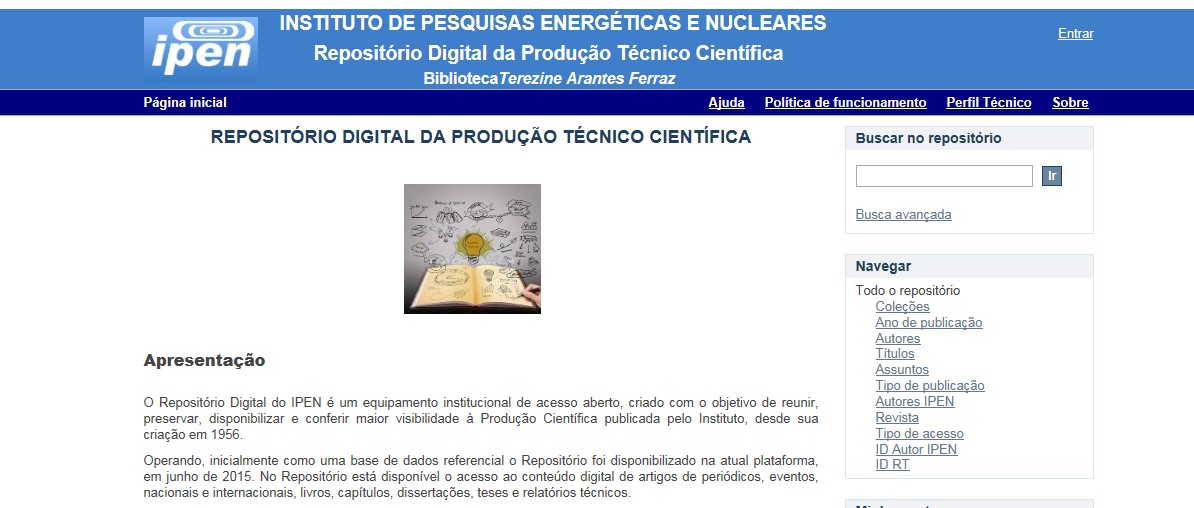
O Repositório Digital do IPEN é um equipamento institucional de acesso aberto, criado com o objetivo de reunir, preservar, disponibilizar e conferir maior visibilidade à Produção Científica publicada pelo Instituto, desde sua criação em 1956.
Operando, inicialmente como uma base de dados referencial o Repositório foi disponibilizado na atual plataforma, em junho de 2015. No Repositório está disponível o acesso ao conteúdo digital de artigos de periódicos, eventos, nacionais e internacionais, livros, capítulos, dissertações, teses e relatórios técnicos.
A elaboração do projeto do RI do IPEN foi iniciado em novembro de 2013, colocado em operação interna em julho de 2014 e disponibilizado na Internet em junho de 2015. Utiliza o software livre Dspace, desenvolvido pelo Massachusetts Institute of Technology (MIT). Para descrição dos metadados adota o padrão Dublin Core. É compatível com o Protocolo de Arquivos Abertos (OAI) permitindo interoperabilidade com repositórios de âmbito nacional e internacional.
O gerenciamento do Repositório está a cargo da Biblioteca do IPEN. Constam neste RI, até o presente momento 20.950 itens que tanto podem ser artigos de periódicos ou de eventos nacionais e internacionais, dissertações e teses, livros, capítulo de livros e relatórios técnicos. Para participar do RI-IPEN é necessário que pelo menos um dos autores tenha vínculo acadêmico ou funcional com o Instituto. Nesta primeira etapa de funcionamento do RI, a coleta das publicações é realizada periodicamente pela equipe da Biblioteca do IPEN, extraindo os dados das bases internacionais tais como a Web of Science, Scopus, INIS, SciElo além de verificar o Currículo Lattes. O RI-IPEN apresenta também um aspecto inovador no seu funcionamento. Por meio de metadados específicos ele está vinculado ao sistema de gerenciamento das atividades do Plano Diretor anual do IPEN (SIGEPI). Com o objetivo de fornecer dados numéricos para a elaboração dos indicadores da Produção Cientifica Institucional, disponibiliza uma tabela estatística registrando em tempo real a inserção de novos itens. Foi criado um metadado que contém um número único para cada integrante da comunidade científica do IPEN. Esse metadado se transformou em um filtro que ao ser acionado apresenta todos os trabalhos de um determinado autor independente das variáveis na forma de citação do seu nome.
