Navegação Periódicos - Resumos por assunto "tomography"
- Página inicial
- →
- IPEN
- →
- Periódicos - Resumos
- →
- Navegação Periódicos - Resumos por assunto
- Sobre
- Perfil Técnico
- Política de funcionamento
- Ajuda
- Apresentação
Navegação Periódicos - Resumos por assunto "tomography"
Itens para a visualização no momento 1-15 de 15
-
. Alternative methods for determining shrinkage in restorative resin composites. Brazilian Oral Research, v. 24, n. Supl. 1, p. 20, 2010.
Palavras-Chave: teeth; biological recovery; resins; shrinkage; tomography; optical properties
. Alternative methods for determining shrinkage in restorative resin composites. Brazilian Oral Research, v. 24, n. Supl. 1, p. 20, 2010. Disponível em: http://repositorio.ipen.br/handle/123456789/8851. Acesso em: $DATA.Como referenciar este itemEsta referência é gerada automaticamente de acordo com as normas do estilo IPEN/SP (ABNT NBR 6023) e recomenda-se uma verificação final e ajustes caso necessário.
-
. Analise comparativa de tomografia por coerência óptica e microdureza para avaliação de desmineralização de esmalte dental humano. Brazilian Oral Research, v. 25, n. Supl. 1, p. 297, 2011.
Palavras-Chave: teeth; enamels; demineralization; tomography; coherent radiation; microhardness; comparative evaluations
. Analise comparativa de tomografia por coerência óptica e microdureza para avaliação de desmineralização de esmalte dental humano. Brazilian Oral Research, v. 25, n. Supl. 1, p. 297, 2011. Disponível em: http://repositorio.ipen.br/handle/123456789/8889. Acesso em: $DATA.Como referenciar este itemEsta referência é gerada automaticamente de acordo com as normas do estilo IPEN/SP (ABNT NBR 6023) e recomenda-se uma verificação final e ajustes caso necessário.
-
. Análise de restaurações em resinas compostas à base de metacrilato e silorano através da Tomografia por Coerência Óptica. Brazilian Oral Research, v. 24, n. Supl. 1, p. 308, 2010.
Palavras-Chave: teeth; biological recovery; resins; methacrylates; tomography; optical properties
. Análise de restaurações em resinas compostas à base de metacrilato e silorano através da Tomografia por Coerência Óptica. Brazilian Oral Research, v. 24, n. Supl. 1, p. 308, 2010. Disponível em: http://repositorio.ipen.br/handle/123456789/8850. Acesso em: $DATA.Como referenciar este itemEsta referência é gerada automaticamente de acordo com as normas do estilo IPEN/SP (ABNT NBR 6023) e recomenda-se uma verificação final e ajustes caso necessário.
-
. Assessment of the optical attenuation coefficient of erored dentin. Lasers in Surgery and Medicine, v. 47, Suppl. 26, p. 32-33, 2015.
Palavras-Chave: coherent radiation; attenuation; dentin; neodymium lasers; fluorine; diagnostic uses; tomography; optical models
. Assessment of the optical attenuation coefficient of erored dentin. Lasers in Surgery and Medicine, v. 47, p. 32-33, 2015. Suppl. 26. Disponível em: http://repositorio.ipen.br/handle/123456789/23867. Acesso em: $DATA.Como referenciar este itemEsta referência é gerada automaticamente de acordo com as normas do estilo IPEN/SP (ABNT NBR 6023) e recomenda-se uma verificação final e ajustes caso necessário.
-
. Assessment to the optical attenuation coefficient of erored dentin. Lasers in Surgery and Medicine, v. 47, Sippl. 26, p. 32-33, 2015.
Palavras-Chave: attenuation; dentin; in vitro; evaluation; tomography; irradiation; neodymium lasers; fluorides; yttrium compounds
. Assessment to the optical attenuation coefficient of erored dentin. Lasers in Surgery and Medicine, v. 47, p. 32-33, 2015. Sippl. 26. Disponível em: http://repositorio.ipen.br/handle/123456789/26467. Acesso em: $DATA.Como referenciar este itemEsta referência é gerada automaticamente de acordo com as normas do estilo IPEN/SP (ABNT NBR 6023) e recomenda-se uma verificação final e ajustes caso necessário.
-
. Avaliação da profundidade de desmineralização da dentina cariada artificialmente através de Tomografia por Coerência Óptica. Brazilian Oral Research, v. 24, n. Supl. 1, p. 120, 2010.
Palavras-Chave: teeth; dentin; caries; demineralization; tomography; optical properties
. Avaliação da profundidade de desmineralização da dentina cariada artificialmente através de Tomografia por Coerência Óptica. Brazilian Oral Research, v. 24, n. Supl. 1, p. 120, 2010. Disponível em: http://repositorio.ipen.br/handle/123456789/8849. Acesso em: $DATA.Como referenciar este itemEsta referência é gerada automaticamente de acordo com as normas do estilo IPEN/SP (ABNT NBR 6023) e recomenda-se uma verificação final e ajustes caso necessário.
-
. Avaliação de resina composta de baixa contração por tomografia de coerência óptica. Brazilian Oral Research, v. 24, n. Supl. 1, p. 344, 2010.
Palavras-Chave: teeth; resins; biological recovery; polymerization; tomography; optical properties
. Avaliação de resina composta de baixa contração por tomografia de coerência óptica. Brazilian Oral Research, v. 24, n. Supl. 1, p. 344, 2010. Disponível em: http://repositorio.ipen.br/handle/123456789/8847. Acesso em: $DATA.Como referenciar este itemEsta referência é gerada automaticamente de acordo com as normas do estilo IPEN/SP (ABNT NBR 6023) e recomenda-se uma verificação final e ajustes caso necessário.
-
. Caracterização de agentes cimentantes resinosos através de Tomografia por Coerência Óptica. Brazilian Oral Research, v. 24, n. Supl. 1, p. 265, 2010.
Palavras-Chave: teeth; tomography; optical properties; resins; fiberglass; prostheses
. Caracterização de agentes cimentantes resinosos através de Tomografia por Coerência Óptica. Brazilian Oral Research, v. 24, n. Supl. 1, p. 265, 2010. Disponível em: http://repositorio.ipen.br/handle/123456789/8853. Acesso em: $DATA.Como referenciar este itemEsta referência é gerada automaticamente de acordo com as normas do estilo IPEN/SP (ABNT NBR 6023) e recomenda-se uma verificação final e ajustes caso necessário.
-
. Emprego da tomografia por coerência óptica na caracterização da interface de pinos estéticos após ensaio de extrusão. Brazilian Oral Research, v. 24, n. Supl. 1, p. 302, 2010.
Palavras-Chave: teeth; tomography; optical properties; morphology; fiberglass; prostheses
. Emprego da tomografia por coerência óptica na caracterização da interface de pinos estéticos após ensaio de extrusão. Brazilian Oral Research, v. 24, n. Supl. 1, p. 302, 2010. Disponível em: http://repositorio.ipen.br/handle/123456789/8852. Acesso em: $DATA.Como referenciar este itemEsta referência é gerada automaticamente de acordo com as normas do estilo IPEN/SP (ABNT NBR 6023) e recomenda-se uma verificação final e ajustes caso necessário.
-
. Imagin carious human dental tissue with three-dimensional optical coherence tomography. Brazilian Dental Journal, v. 15, Special issue, p. 79-79, 2004. Abstract: Optical Coherence Tomography (OCT) used in this study, is a new non invasive optical detection technique. The OCT system is based on a Michelson interferometer, that generates a crosssectional image of the teeth with resolution up to 2 microns. The buccal surface from the third molar teeth was used to induce caries like lesions. This surface was coated with an acid resistant nail varnish except a small window. The pH demineralizationremineralization cycling model was used to produce the lesions. This cycle was repeated for 9 days and remained in the remineralizing solution for 2 days. The OCT system was implemented by using an ultrashort pulse laser (Ti:Al2O3@830nm) with 50fs of pulse width and average power of 80mW. The laser beam was focused into the teeth providing a lateral resolution of 10 microns. Image was produced with a lateral and axial scans steps of 10 microns. After analyzing the surface by OCT it was possible to produce a tomogram of dentine-enamel junction and it was compared with the histological image. This OCT system accurately depicts dental tissue and it was able to detect early caries in its structure, providing a powerful contactless high resolution 3D images of lesions.
Palavras-Chave: teeth; bone tissues; microstructure; caries; three-dimensional calculations; interferometry; optical properties; tomography
. Imagin carious human dental tissue with three-dimensional optical coherence tomography. Brazilian Dental Journal, v. 15, p. 79-79, 2004. Special issue. Disponível em: http://repositorio.ipen.br/handle/123456789/32735. Acesso em: $DATA.Como referenciar este itemEsta referência é gerada automaticamente de acordo com as normas do estilo IPEN/SP (ABNT NBR 6023) e recomenda-se uma verificação final e ajustes caso necessário.
-
. New method for depth analysis of Y-TZP t-m phase transformation. Dental Materials, v. 33, suppl. 1, p. e6-e6, 2017. DOI: 10.1016/j.dental.2017.08.009 Abstract: Purpose/aim: The aim of this studywas to validate the optical coherence tomography (OCT) as a nondestructive method of analysis to evaluate the depth of tetragonal to monoclinic (t-m) transformed zone and to calculate the kinetics of phase transformation of a monolithic Y-TZP after hydrothermal aging. Specifically, to compare the activation energy of t-m transformation calculated by the depth of the transformed zone using scanning electron microscopy (SEM) and OCT. Materials and methods: Fully sintered (1450 ◦C/2 h) discs of dentalY-TZP (LAVAPLUS, 3M-ESPE)were aged in hydrothermal pressurized reactor to follow the phase transformation kinetics at 120 to 150 ◦C. Four samples per aging time were analyzed by OCT (OCP930SR, Thorlabs Inc.), = 930 nm, spectral bandwidth (FWHM) of 100 nm, nominal resolution of 6 m (lateral and axial) in air, declared digital resolution 3.09 m (axial). Three areas of 3mm (lateral) were observed to calculate the phase transformation depth (Image J). X-ray diffraction analysis (XRD) were performed, Cu-K , 20◦ to 80◦, 2 . The data were refined using the Rietveld method (GSAS). The transversal section of one specimen of each group was submitted to backscattered SEM analysis to calculate the phase transformation depth (Image J). The speed of the transformation zone front was determined plotting the phase transformation depth versus aging time. Results: XRD results indicated that Y-TZP that 66% is the maximum value of monoclinic phase concentration for all aged Y-TZP. The activation energy for the monolithic Y-TZP was 107.53 kJ/mol. One year and 5 years of hydrothermal aging at 37 ◦C will present approximately 4.21% and 15% of monoclinic phase, respectively. The comparison of the depth of the transformed zone using SEM and OCT were similar, showing a linear behavior and providing information that the opaque layer observed by OCT is related to the depth of the transformed zone (Fig. 1), any difference among the results could be a result of the refraction index correction. The energy of activation calculated by SEM and OCT were 114 kJ/mol and 100 kJ/mol, respectively. The speed calculated for the phase transformation into the bulk of the transformed zone estimated for 37 ◦C was 0.04 m/year (SEM) and 0.16 m/year (OCT). Conclusions: The results indicate that activation energy values determined by SEM and OCT observations were similar allowing the use of the OCT as a tool for monolithic Y-TZP t-m phase transformation kinetic evaluation. Moreover, OCT method has the advantage of a shorter analysis time, without the need of sample preparation steps.
Palavras-Chave: crystal-phase transformations; tomography; optical modes; coherence length; scanning electron microscopy; measuring methods
. New method for depth analysis of Y-TZP t-m phase transformation. Dental Materials, v. 33, p. e6-e6, 2017. suppl. 1, DOI: 10.1016/j.dental.2017.08.009. Disponível em: http://repositorio.ipen.br/handle/123456789/28641. Acesso em: $DATA.Como referenciar este itemEsta referência é gerada automaticamente de acordo com as normas do estilo IPEN/SP (ABNT NBR 6023) e recomenda-se uma verificação final e ajustes caso necessário.
-
. Optical and histological evaluation of human tendon tissue sterilized by ionizing radiation. Regenerative Research, v. 7, n. 1, p. 122-122, 2018. Abstract: Sterilization by irradiation is a technique that is used by tissue banks aiming to eliminate contamination of human allografts, being a safe method, free of residue and used as final sterilization. After the tissue procurement, these undergo a series of processing stages and then are packaged and preserved by freezing. Despite aseptic care of the material those may be subjected to sterilization in the final packing by ionizing radiation, raising the security level of sterility of the tissue. The aim of this study was to evaluate the effects of application of ionizing radiation, produced by 60Co source in human tendons preprocessed (A-alcohol + antibiotic; B- H2O2 + ultrasound) obtained through collaboration with tissue banks and preserved by freezing in -80° C, the radiation absorbed doses in processing were 12.5, 15 and 25 kGy, each one with their corresponding non-irradiated control, to examine possible structural or morphological alterations. The irradiated samples and their controls were analyzed by means of optical coherence tomography (OCT) and optical coherence tomography polarization sensitive (PS-OCT), and histological tests had been stained with hematoxylin-eosin (HE). According to the results the tissue processed with alcohol/antibiotic in conjunction with irradiation proved to be the most effective.
Palavras-Chave: absorbed radiation doses; alcohols; animal tissues; antibiotics; cobalt 60; freezing; histological techniques; ionizing radiations; radiation effects; sterilization; tendons; tomography
. Optical and histological evaluation of human tendon tissue sterilized by ionizing radiation. Regenerative Research, v. 7, n. 1, p. 122-122, 2018. Disponível em: http://repositorio.ipen.br/handle/123456789/29715. Acesso em: $DATA.Como referenciar este itemEsta referência é gerada automaticamente de acordo com as normas do estilo IPEN/SP (ABNT NBR 6023) e recomenda-se uma verificação final e ajustes caso necessário.
-
. Optical coherence tomography as a method for quantitative skin evaluation in systemic sclerosis. Annals of the Rheumatic Diseases, v. 77, n. 03, p. 465-466, 2018. DOI: 10.1136/annrheumdis-2016-210875
Palavras-Chave: rheumatic diseases; diagnostic techniques; optics; tomography; skin; fibrosis
. Optical coherence tomography as a method for quantitative skin evaluation in systemic sclerosis. Annals of the Rheumatic Diseases, v. 77, n. 03, p. 465-466, 2018. DOI: 10.1136/annrheumdis-2016-210875. Disponível em: http://repositorio.ipen.br/handle/123456789/27386. Acesso em: $DATA.Como referenciar este itemEsta referência é gerada automaticamente de acordo com as normas do estilo IPEN/SP (ABNT NBR 6023) e recomenda-se uma verificação final e ajustes caso necessário.
-
. Optical coherence tomography assessment of chronological ageing: A population study. Journal of Investigate Dermatology, v. 135, n. 8, p. S2, 2015.
Palavras-Chave: tomography; aging; in vivo; clinical trials; skin; roughness; morphological changes; epidermis
. Optical coherence tomography assessment of chronological ageing: A population study. Journal of Investigate Dermatology, v. 135, n. 8, p. S2, 2015. Disponível em: http://repositorio.ipen.br/handle/123456789/25849. Acesso em: $DATA.Como referenciar este itemEsta referência é gerada automaticamente de acordo com as normas do estilo IPEN/SP (ABNT NBR 6023) e recomenda-se uma verificação final e ajustes caso necessário.
-
. Special section on the 8th World Congress on Industrial Process Tomography (WCIPT8). Measurement Science and Technology, v. 29, n. 060101, 2018. DOI: 10.1088/1361-6501/aabb44 Abstract: Multiphase flow systems are widely used in the industrial production of polymers, minerals, pharmaceuticals and food processing, among others. Industrial process tomography (IPT) may contribute to increase the production and quality control of the means of production or the industrial product itself. There has been a remarkable development in IPT technology over the past two decades, a fact demonstrated by the quality and innovation of the works presented at the 8th World Congress on Industrial Process Tomography (WCIPT8). This scientific meeting, held in Foz do Iguaçu, Brazil, in September 2016, was crowned by a high scientific and technological level within the natural beauty of the Iguaçu Falls. New sensor technologies and the processing of electronic signals have been continuously under development, and these themes were discussed in the WCIPT8. These new technologies are being implemented in the new IPT versions and, as a corollary, some improvement in the sensitivity of what may be measured with modern IPTs, with the desired accuracy, has been observed. In addition, the miniaturization of sensors and the use of nano-technology push these limits further. Further, thanks to the advancement of modern computers, sophisticated online image reconstruction algorithms are now available and have been incorporated into modern IPT allowing in-situ and on-line data processing. A large number of IPT measurement principles have been and are still under development: electrical methods, such as the measurement of capacitance, inductance and resistance, optical and radiation-based methods, ranging from infrared, microwave, x-rays, gamma rays and even neutrons, magnetic resonance, ultrasound and acoustic methods, to mention a few. The sensor technology for a specific application is primarily selected to achieve sensitivity to a physical property which differs for each of the components of the process, e.g. density or electrical permittivity. For the measurement or imaging of more than two components, multi-modality systems are often employed, either by measuring with one principle at several wavelengths or energies, or by combining several independent sensor principles. IPT is inherently interdisciplinary, so that R&D requires skills in each of the engineering processes (chemical, combustion, pharmaceutical, etc), physics and electronic engineering for the sensor system, plus mathematics and computer science for data processing algorithms. This special issue of Measurement Science and Technology presents innovative papers on IPT technologies, presented at the 8th World Congress on Industrial Process Tomography (WCIPT8), promoted by the International Society on Industrial Process Tomography (ISIPT) and organized by the Nuclear and Energy Research Institute (IPEN) and the Federal University of Technology, Parana. We hope that all readers of this Special Section may find not only interesting information, but also data that is useful for their scientific work.
Palavras-Chave: industrial radiography; tomography; industry; appropriate technology; meetings
. Special section on the 8th World Congress on Industrial Process Tomography (WCIPT8). Measurement Science and Technology, v. 29, n. 060101, 2018. DOI: 10.1088/1361-6501/aabb44. Disponível em: http://repositorio.ipen.br/handle/123456789/28955. Acesso em: $DATA.Como referenciar este itemEsta referência é gerada automaticamente de acordo com as normas do estilo IPEN/SP (ABNT NBR 6023) e recomenda-se uma verificação final e ajustes caso necessário.
Itens para a visualização no momento 1-15 de 15
Buscar no repositório
Navegar
Minha conta
Visualizar
A pesquisa no RD utiliza os recursos de busca da maioria das bases de dados. No entanto algumas dicas podem auxiliar para obter um resultado mais pertinente.
✔ É possível efetuar a busca de um autor ou um termo em todo o RD, por meio do Buscar no Repositório , isto é, o termo solicitado será localizado em qualquer campo do RD. No entanto esse tipo de pesquisa não é recomendada a não ser que se deseje um resultado amplo e generalizado.
✔ A pesquisa apresentará melhor resultado selecionando um dos filtros disponíveis em Navegar
✔ Os filtros disponíveis em Navegar tais como: Coleções, Ano de publicação, Títulos, Assuntos, Autores, Revista, Tipo de publicação são autoexplicativos. O filtro, Autores IPEN apresenta uma relação com os autores vinculados ao IPEN; o ID Autor IPEN diz respeito ao número único de identificação de cada autor constante no RD e sob o qual estão agrupados todos os seus trabalhos independente das variáveis do seu nome; Tipo de acesso diz respeito à acessibilidade do documento, isto é , sujeito as leis de direitos autorais, ID RT apresenta a relação dos relatórios técnicos, restritos para consulta das comunidades indicadas.

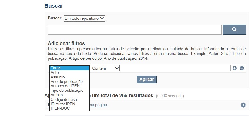
A opção Busca avançada utiliza os conectores da lógica boleana, é o melhor recurso para combinar chaves de busca e obter documentos relevantes à sua pesquisa, utilize os filtros apresentados na caixa de seleção para refinar o resultado de busca. Pode-se adicionar vários filtros a uma mesma busca.
Exemplo:
Buscar os artigos apresentados em um evento internacional de 2015, sobre loss of coolant, do autor Maprelian.
Autor: Maprelian
Título: loss of coolant
Tipo de publicação: Texto completo de evento
Ano de publicação: 2015

✔ Para indexação dos documentos é utilizado o Thesaurus do INIS, especializado na área nuclear e utilizado em todos os países membros da International Atomic Energy Agency – IAEA , por esse motivo, utilize os termos de busca de assunto em inglês; isto não exclui a busca livre por palavras, apenas o resultado pode não ser tão relevante ou pertinente.
✔ 95% do RD apresenta o texto completo do documento com livre acesso, para aqueles que apresentam o ![]() significa que e o documento está sujeito as leis de direitos autorais, solicita-se nesses casos contatar a Biblioteca do IPEN,
bibl@ipen.br
.
significa que e o documento está sujeito as leis de direitos autorais, solicita-se nesses casos contatar a Biblioteca do IPEN,
bibl@ipen.br
.
✔ Ao efetuar a busca por um autor o RD apresentará uma relação de todos os trabalhos depositados no RD. No lado direito da tela são apresentados os coautores com o número de trabalhos produzidos em conjunto bem como os assuntos abordados e os respectivos anos de publicação agrupados.
✔ O RD disponibiliza um quadro estatístico de produtividade, onde é possível visualizar o número dos trabalhos agrupados por tipo de coleção, a medida que estão sendo depositados no RD.
✔ Na página inicial nas referências são sinalizados todos os autores IPEN, ao clicar nesse símbolo ![]() será aberta uma nova página correspondente à aquele autor – trata-se da página do pesquisador.
será aberta uma nova página correspondente à aquele autor – trata-se da página do pesquisador.
✔ Na página do pesquisador, é possível verificar, as variações do nome, a relação de todos os trabalhos com texto completo bem como um quadro resumo numérico; há links para o Currículo Lattes e o Google Acadêmico ( quando esse for informado).
ATENÇÃO!
ESTE TEXTO "AJUDA" ESTÁ SUJEITO A ATUALIZAÇÕES CONSTANTES, A MEDIDA QUE NOVAS FUNCIONALIDADES E RECURSOS DE BUSCA FOREM SENDO DESENVOLVIDOS PELAS EQUIPES DA BIBLIOTECA E DA INFORMÁTICA.
O gerenciamento do Repositório está a cargo da Biblioteca do IPEN. Constam neste RI, até o presente momento 20.950 itens que tanto podem ser artigos de periódicos ou de eventos nacionais e internacionais, dissertações e teses, livros, capítulo de livros e relatórios técnicos. Para participar do RI-IPEN é necessário que pelo menos um dos autores tenha vínculo acadêmico ou funcional com o Instituto. Nesta primeira etapa de funcionamento do RI, a coleta das publicações é realizada periodicamente pela equipe da Biblioteca do IPEN, extraindo os dados das bases internacionais tais como a Web of Science, Scopus, INIS, SciElo além de verificar o Currículo Lattes. O RI-IPEN apresenta também um aspecto inovador no seu funcionamento. Por meio de metadados específicos ele está vinculado ao sistema de gerenciamento das atividades do Plano Diretor anual do IPEN (SIGEPI). Com o objetivo de fornecer dados numéricos para a elaboração dos indicadores da Produção Cientifica Institucional, disponibiliza uma tabela estatística registrando em tempo real a inserção de novos itens. Foi criado um metadado que contém um número único para cada integrante da comunidade científica do IPEN. Esse metadado se transformou em um filtro que ao ser acionado apresenta todos os trabalhos de um determinado autor independente das variáveis na forma de citação do seu nome.
A elaboração do projeto do RI do IPEN foi iniciado em novembro de 2013, colocado em operação interna em julho de 2014 e disponibilizado na Internet em junho de 2015. Utiliza o software livre Dspace, desenvolvido pelo Massachusetts Institute of Technology (MIT). Para descrição dos metadados adota o padrão Dublin Core. É compatível com o Protocolo de Arquivos Abertos (OAI) permitindo interoperabilidade com repositórios de âmbito nacional e internacional.
1. Portaria IPEN-CNEN/SP nº 387, que estabeleceu os princípios que nortearam a criação do RDI, clique aqui.
2. A experiência do Instituto de Pesquisas Energéticas e Nucleares (IPEN-CNEN/SP) na criação de um Repositório Digital Institucional – RDI, clique aqui.
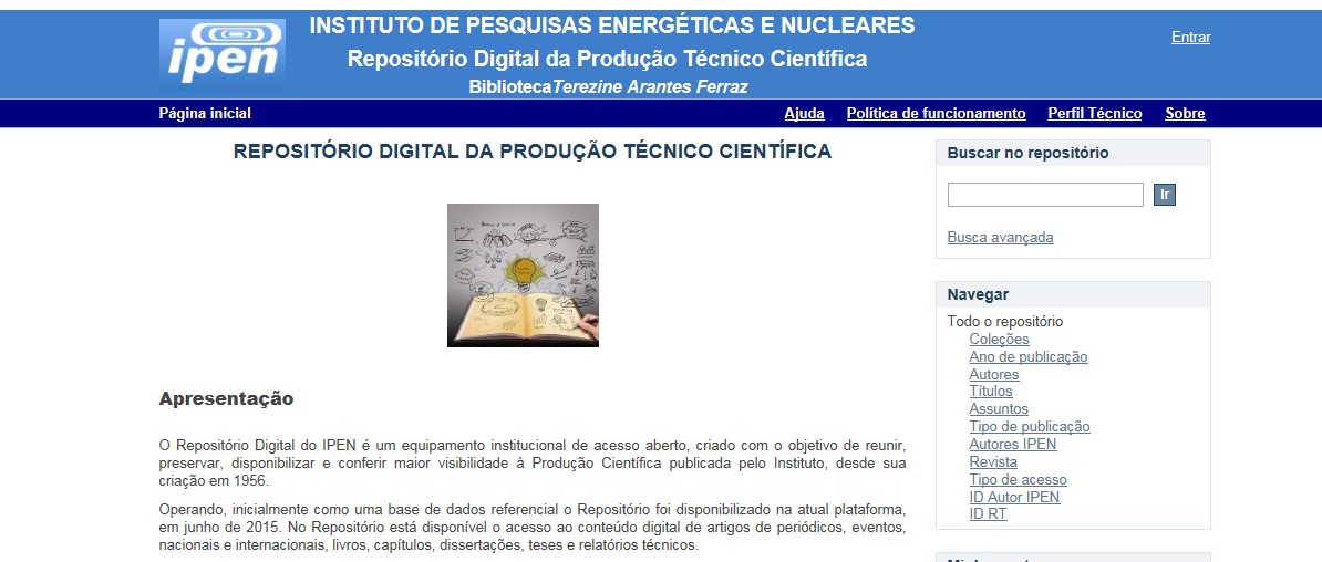
O Repositório Digital do IPEN é um equipamento institucional de acesso aberto, criado com o objetivo de reunir, preservar, disponibilizar e conferir maior visibilidade à Produção Científica publicada pelo Instituto, desde sua criação em 1956.
Operando, inicialmente como uma base de dados referencial o Repositório foi disponibilizado na atual plataforma, em junho de 2015. No Repositório está disponível o acesso ao conteúdo digital de artigos de periódicos, eventos, nacionais e internacionais, livros, capítulos, dissertações, teses e relatórios técnicos.
A elaboração do projeto do RI do IPEN foi iniciado em novembro de 2013, colocado em operação interna em julho de 2014 e disponibilizado na Internet em junho de 2015. Utiliza o software livre Dspace, desenvolvido pelo Massachusetts Institute of Technology (MIT). Para descrição dos metadados adota o padrão Dublin Core. É compatível com o Protocolo de Arquivos Abertos (OAI) permitindo interoperabilidade com repositórios de âmbito nacional e internacional.
O gerenciamento do Repositório está a cargo da Biblioteca do IPEN. Constam neste RI, até o presente momento 20.950 itens que tanto podem ser artigos de periódicos ou de eventos nacionais e internacionais, dissertações e teses, livros, capítulo de livros e relatórios técnicos. Para participar do RI-IPEN é necessário que pelo menos um dos autores tenha vínculo acadêmico ou funcional com o Instituto. Nesta primeira etapa de funcionamento do RI, a coleta das publicações é realizada periodicamente pela equipe da Biblioteca do IPEN, extraindo os dados das bases internacionais tais como a Web of Science, Scopus, INIS, SciElo além de verificar o Currículo Lattes. O RI-IPEN apresenta também um aspecto inovador no seu funcionamento. Por meio de metadados específicos ele está vinculado ao sistema de gerenciamento das atividades do Plano Diretor anual do IPEN (SIGEPI). Com o objetivo de fornecer dados numéricos para a elaboração dos indicadores da Produção Cientifica Institucional, disponibiliza uma tabela estatística registrando em tempo real a inserção de novos itens. Foi criado um metadado que contém um número único para cada integrante da comunidade científica do IPEN. Esse metadado se transformou em um filtro que ao ser acionado apresenta todos os trabalhos de um determinado autor independente das variáveis na forma de citação do seu nome.
